
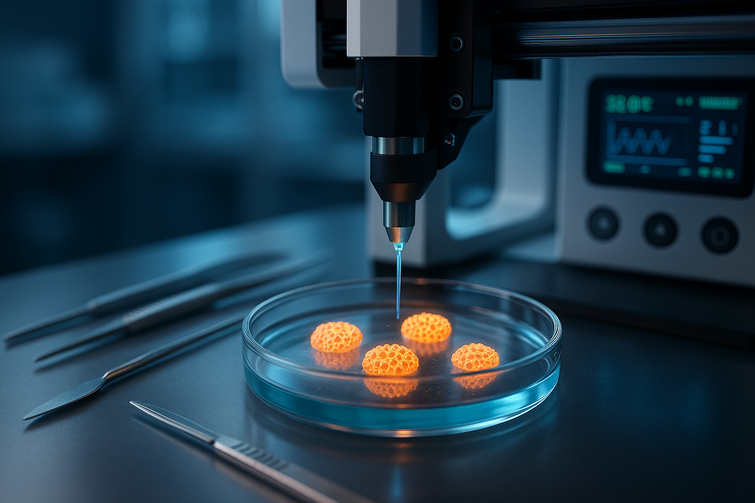

What if scientists could use a 3D printer to create structures that help insulin-producing cells survive longer and respond to changes in blood sugar?
For people living with type 1 diabetes, the possibility is significant. Current treatments can manage blood glucose effectively, but they usually don’t restore the body’s ability to produce its own insulin.
Researchers are now exploring a different approach: creating living, three-dimensional structures that support pancreatic islets, the tiny clusters of cells that produce insulin and other hormones.
In June 2025, an international research team presented an experimental bioprinting method at the European Society for Organ Transplantation (ESOT) Congress. Their work showed that human pancreatic islets could remain alive and functional for three weeks inside a specially formulated 3D-printed material.
The research has since undergone peer review. In September 2026, scientists published their detailed findings in Acta Biomaterialia, providing new evidence about the technique’s potential for future cell-replacement therapies.
The study found that more than 85% of the islets remained viable and continued responding to glucose under laboratory conditions.
However, the printed structures have not been demonstrated to cure type 1 diabetes in humans. Their effectiveness after transplantation, long-term safety, and ability to withstand immune attack remain unanswered questions.
Here’s what the research actually shows, why it matters, and what scientists must accomplish before the technology could become a treatment.
Understanding Type 1 Diabetes: Why Insulin-Producing Cells Matter
Type 1 diabetes is a chronic autoimmune condition in which the immune system attacks pancreatic beta cells.
These specialized cells produce insulin, a hormone that helps glucose move from the bloodstream into cells, where it can be used or stored.
When enough beta cells are destroyed, the pancreas can no longer produce the insulin the body needs.
Without adequate insulin, blood glucose rises, and serious complications can develop, including diabetic ketoacidosis, a potentially life-threatening emergency.
People living with type 1 diabetes therefore require insulin replacement.
Treatment commonly involves insulin injections or continuous delivery through an insulin pump. Continuous glucose monitors and automated insulin-delivery systems can help improve blood glucose management.
These technologies have transformed diabetes care, but they do not replace the lost pancreatic beta cells.
That is why scientists are investigating treatments that could restore some degree of natural insulin production.
One possibility is pancreatic islet transplantation.
What Are Pancreatic Islets?
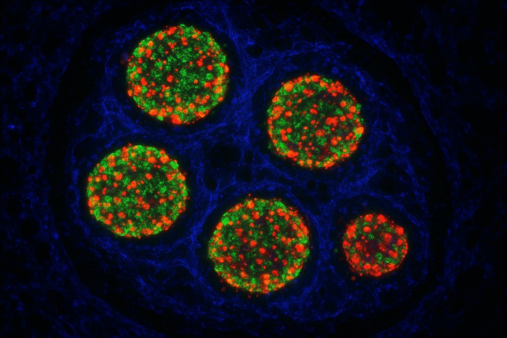
Pancreatic islets, also called the islets of Langerhans, are small groups of hormone-producing cells located throughout the pancreas.
They contain several cell types, including:
- Beta cells, which produce insulin and help lower blood glucose.
- Alpha cells, which produce glucagon and help raise blood glucose when necessary.
- Delta cells, which release somatostatin and help regulate hormone secretion.
Together, these cells help maintain normal glucose levels.
In people without diabetes, beta cells respond to increasing glucose concentrations by releasing insulin.
Scientists hope that transplanting healthy, functioning islets could restore some of this natural responsiveness in people with type 1 diabetes.
But maintaining transplanted islets is challenging.
Why Islets Often Struggle After Transplantation
During isolation and transplantation, islets lose much of the supporting environment they normally experience inside the pancreas.
They may also face inadequate oxygen delivery, inflammation, stress, and immune attack.
These problems can reduce the number of surviving cells and impair their ability to release insulin.
Even if transplanted islets function well initially, sustaining their activity over time remains difficult.
Researchers are therefore exploring ways to recreate the pancreas’s supportive environment.
This is where 3D bioprinting comes in.
What Is 3D Bioprinting?
Traditional 3D printing builds objects by depositing layers of plastic, resin, metal, or other materials according to a computer-generated design.
3D bioprinting uses a related principle but incorporates biological materials, often including living cells.
Instead of producing an ordinary manufactured object, a bioprinter deposits carefully formulated biological materials into three-dimensional arrangements.
The material used during printing is commonly called bioink.
Bioinks may contain living cells, hydrogels, proteins, and other components designed to support cellular survival.
In pancreatic research, the goal is not merely to print the shape of a pancreas.
Scientists want to create an environment in which insulin-producing cells can remain healthy, receive nutrients, and respond appropriately to glucose.
An effective bioink must therefore balance several competing requirements.
It needs to be stable enough to hold a structure’s shape while allowing sufficient transport of oxygen, nutrients, glucose, and insulin.
It must also be gentle enough not to damage living cells during printing.
These requirements make bioprinting considerably more complicated than ordinary 3D printing.
The 2025 Breakthrough: Printing Functional Human Islets
The research highlighted in your original July 2025 article came from an international collaboration involving scientists at Wake Forest University School of Medicine and other institutions.
The findings were presented at the ESOT Congress in June 2025.
Researchers reported developing a bioink containing two principal ingredients:
- Alginate, a hydrogel-forming material derived from seaweed.
- Decellularized extracellular matrix (dECM) obtained from human pancreatic tissue.
The team embedded human pancreatic islets in this material and used extrusion-based bioprinting to form three-dimensional structures.
Early results suggested that the printed islets could remain viable and continue secreting insulin in response to glucose for up to three weeks.
The researchers also investigated how to maintain islet function when cells were packed into relatively dense printed structures.
This mattered because high cell density can make it more difficult for oxygen and nutrients to reach every cell.
The initial findings were encouraging, but they were presented at a scientific conference.
The full peer-reviewed study subsequently provided a more detailed evaluation of the technique.
September 2026: The Research Is Published in Acta Biomaterialia
In September 2026, researchers led by Wonwoo Jeong and Quentin Perrier published:
The study appeared in Acta Biomaterialia, volume 222, pages 327–339.
The research explored whether human pancreatic islets could remain structurally stable and functional when embedded in an optimized, pancreas-specific bioink.
Unlike early experiments that focus on small numbers of cells, the researchers also evaluated a printing system intended to support future manufacturing at clinically relevant scales.
How the Scientists Created the Printed Islets
The team developed an alginate-based bioink enriched with human pancreatic dECM.
The extracellular matrix is a network of proteins and other molecules that surrounds cells within tissues.
It provides physical support and can influence cell behavior.
To produce dECM, researchers remove cellular material from donated tissue, leaving behind components of its supporting structure.
By incorporating pancreatic dECM into the bioink, the researchers attempted to recreate some features of the natural pancreatic environment.
They also optimized the extrusion-printing process to reduce mechanical stress on the delicate human islets.
These adjustments helped maintain the printed material’s structural integrity while supporting the exchange of important molecules.
The result was a collection of living human islets embedded within a three-dimensional hydrogel-based structure.
What Did the 2026 Study Actually Find?
The peer-reviewed research reported several important laboratory findings.
1. More Than 85% Islet Viability
The researchers observed viability above 85% in the printed human islet constructs.
Viability refers to the proportion of cells remaining alive under the tested conditions.
Maintaining high viability matters because pancreatic islets can be damaged during isolation, handling, and printing.
A bioprinting process that preserves more living cells may improve the prospects for future transplantation.
However, laboratory viability does not establish survival after implantation in a person.
2. Insulin Secretion Continued for 21 Days
The printed islets continued to respond to glucose during a 21-day laboratory culture period.
When glucose concentrations increased, the cells demonstrated glucose-stimulated insulin secretion.
This is particularly important because insulin release in response to glucose is a fundamental function of pancreatic beta cells.
The researchers compared the printed constructs with free islets maintained under conventional culture conditions.
The free-islet controls showed a significant decline in function over time, while the printed constructs maintained their glucose-responsive activity.
This suggests the 3D environment may have protected the islets from some culture-related stress.
It does not mean the printed islets performed identically to a healthy human pancreas.
3. Function at High Islet Density
The researchers tested a packing density of up to 10,000 islet equivalents per milliliter.
An islet equivalent is a standardized measurement used to compare islet preparations of different sizes.
High-density printing matters because effective cell-replacement therapies may require large numbers of functioning insulin-producing cells.
The printed structures maintained viability and glucose-responsive insulin secretion at the tested density.
This is an encouraging manufacturing finding, although reliably producing clinical-scale constructs remains a separate challenge.
4. The Bioink Supported Structural Stability
The printed material maintained its three-dimensional structure during laboratory culture.
The researchers also designed the hydrogel to allow the transport of nutrients and other important molecules.
These characteristics matter because transplanted cells need a suitable physical environment and access to nutrients to survive.
The researchers proposed that the supportive dECM-rich environment contributed to the improved performance.
But identifying the precise biological mechanisms behind these improvements will require further investigation.
What Is the Difference Between Printed Islets and a Printed Pancreas?
One common misunderstanding is that scientists have already printed an entire functional human pancreas.
This study did not demonstrate that.
The researchers used existing human pancreatic islets and embedded them within a specially engineered material.
The result was a tissue-like construct containing functioning islet cells.
It was not a complete pancreas with the complex blood vessels, ducts, nerves, and multiple tissue structures found in the human organ.
The research also did not demonstrate that the bioprinter could create unlimited new islets from ordinary materials.
The living islet cells still had to come from a biological source.
This distinction matters because donor availability remains a major limitation of islet transplantation.
Bioprinting may help preserve and organize these cells, but it does not automatically solve the shortage of suitable donor tissue.
Why Could This Matter for Future Diabetes Treatment?
A successful islet-replacement therapy could potentially restore some natural insulin secretion.
For people with type 1 diabetes, that could mean more stable blood glucose and, in some cases, less dependence on injected insulin.
But for such a treatment to work, transplanted cells must meet several requirements.
They need to survive implantation, receive adequate oxygen and nutrients, respond appropriately to glucose, and avoid destructive immune responses.
The 2026 bioprinting study addresses parts of this challenge by investigating a supportive environment for islet survival and function.
It does not yet solve all of them.
One potential benefit of engineered tissue constructs is the possibility of designing implantation sites other than the liver, where conventional donor-islet infusion is often performed.
Subcutaneous implantation, for example, has attracted interest because a device placed beneath the skin may be more accessible for monitoring or retrieval.
However, placing cells under the skin creates additional challenges involving oxygen delivery, blood vessel formation, and immune protection.
The 2026 study does not establish that subcutaneous transplantation of its printed constructs is safe or effective in humans.
How Does This Compare With Existing Islet Transplantation?
Islet transplantation is not an entirely new treatment concept.
In June 2023, the U.S. Food and Drug Administration approved Lantidra (donislecel), a donor-derived pancreatic islet cellular therapy.
It is intended for a narrowly defined group of adults with type 1 diabetes who experience repeated severe hypoglycemia despite intensive diabetes management and education.
The therapy uses islets from deceased donors and delivers them to the liver through the hepatic portal vein.
In the FDA-evaluated studies, 21 of 30 participants achieved insulin independence for at least one year.
However, the treatment requires immunosuppression, and serious adverse reactions can occur.
That means it is not a routine replacement for insulin treatment in the general type 1 diabetes population.
Read the FDA announcement about Lantidra.
The experimental 3D-bioprinting approach has a different goal.
Rather than simply infusing isolated islets, it aims to provide cells with an engineered environment that may support their survival and function.
Whether that approach eventually offers advantages over established transplantation techniques remains to be determined through animal studies and human clinical trials.
What Other Research Has Emerged by 2026?
Islet replacement research has advanced along several paths.
Some teams are investigating better protective materials, while others are exploring stem-cell-derived beta cells or genetically engineered donor cells.
These approaches are related but not interchangeable.
3D-Printed Devices With Blood Vessel Support
A study published in Biomaterials in April 2025 described an experimental 3D-printed device containing stem-cell-derived beta-like cells and microvascular fragments.
The researchers investigated whether a printed protective structure and components that support blood vessel formation could improve cell survival after implantation.
In a mouse model, the approach reduced high blood glucose in a subset of treated animals.
The results suggested potential benefits from combining islet engineering with vascular support.
However, the research was preclinical and did not demonstrate effectiveness in humans.
Read the study in Biomaterials.
Progress Toward Avoiding Immune Suppression
Another important research direction involves modifying transplanted cells so the recipient’s immune system is less likely to attack them.
In 2025, a study in The New England Journal of Medicine described early clinical research in which genetically modified donor pancreatic islet cells were transplanted into a person with type 1 diabetes without conventional immunosuppression.
The transplanted cells showed evidence of survival and insulin production during follow-up.
These early findings were encouraging, but they were based on a very small clinical experience and did not establish a generally available treatment.
Importantly, this was not the same technology as the 3D-bioprinted islet constructs reported in Acta Biomaterialia.
The two research directions may eventually complement one another, but that remains speculative.
Read the research announcement and publication context.
A Broader View From a 2026 Scientific Review
A May 2026 review in the Annual Review of Biomedical Engineering examined the evolution of islet encapsulation technologies.
The review discussed how biomaterials, device design, cell sourcing, and immune protection have shaped efforts to develop more effective islet-replacement therapies.
Its broader conclusion reinforces an important point: maintaining insulin-producing cells is only one part of a successful transplant strategy.
Long-term survival, immune compatibility, oxygen delivery, and manufacturing consistency must also be addressed.
Read the 2026 review on islet encapsulation.
The Biggest Challenges Before 3D-Bioprinted Islets Reach Patients
The laboratory findings are promising, but several major barriers remain.
1. Immune Rejection
Type 1 diabetes involves autoimmune destruction of beta cells.
Even if transplanted islets function well initially, immune responses may damage them.
Donor-derived cells can also trigger rejection because the recipient’s immune system recognizes them as foreign.
Some biomaterials may help create a protective barrier, but the 2026 bioprinting study did not establish that its constructs can prevent immune rejection in humans.
2. Oxygen and Blood Supply
Islets require oxygen and nutrients to survive.
Inside the body, thick or densely packed cell structures may not receive sufficient oxygen unless an adequate blood supply develops nearby.
Poor oxygenation can damage cells and reduce insulin production.
Researchers therefore need to establish whether printed islet structures can function after implantation.
3. Long-Term Stability
The 2026 study demonstrated glucose-responsive insulin secretion for 21 days in laboratory culture.
That is a meaningful result, but a useful clinical treatment would likely need to function for much longer.
Scientists must evaluate how printed structures behave over months or years, including whether the material degrades or triggers inflammation.
4. Sourcing Enough Cells
The technology currently relies on obtaining functional human islets.
Donor availability remains limited.
Stem-cell-derived islets could potentially provide an alternative source, but these cells introduce their own manufacturing, safety, and quality-control challenges.
5. Manufacturing and Regulation
Clinical applications require reproducible manufacturing methods.
Printed constructs must contain sufficient viable cells, maintain consistent function, and meet strict quality requirements.
Future regulatory evaluation would also need to address risks associated with the living cells and the biomaterials used to support them.
Could 3D Bioprinting Eliminate the Need for Insulin Injections?
Possibly for some patients in the future, but it is too early to draw that conclusion from this research.
The 2026 study showed that bioprinted human islets could remain alive and release insulin during laboratory culture.
It did not demonstrate that the constructs restored normal blood glucose in a person with type 1 diabetes.
A future implant would need to produce enough insulin at the right times, respond appropriately to changing glucose levels, and remain functional over an extended period.
Researchers would also need to establish that the procedure’s benefits outweigh its risks.
Existing islet transplantation has already shown that cell replacement can sometimes produce insulin independence in carefully selected patients.
But it requires specialized treatment and may involve significant risks.
Bioprinting represents a potential way to improve future cell-replacement technologies, not a currently established substitute for insulin therapy.
Does This Mean a Cure for Type 1 Diabetes Is Close?
The word cure requires careful interpretation.
Type 1 diabetes is driven by an autoimmune process that destroys insulin-producing beta cells.
Restoring insulin secretion through transplanted cells could provide substantial benefits, but it would not necessarily eliminate the underlying autoimmune tendency.
A durable treatment would need to preserve functioning beta cells and prevent their destruction.
Some researchers distinguish between a biological cure that resolves the underlying disease process and a functional cure that allows normal glucose regulation without ongoing external insulin.
Both are ambitious research goals.
The current bioprinting findings do not establish either outcome in patients.
What Should People With Type 1 Diabetes Do Now?
The most important practical message is to continue existing diabetes treatment as prescribed.
Insulin remains essential for people with type 1 diabetes unless a qualified specialist determines otherwise.
Modern insulin pumps, continuous glucose monitoring, diabetes education, and individualized medical care can help reduce complications and improve quality of life.
People who experience severe hypoglycemia despite intensive treatment may benefit from discussion with a specialist about advanced treatment options.
However, islet transplantation and experimental cell therapies are not appropriate for every patient.
Anyone interested in participating in research should consult an established diabetes center and use a recognized clinical trial registry.
Search registered type 1 diabetes clinical trials.
Research announcements should not be interpreted as evidence that a new treatment is already available.
What Happens Next in 3D-Bioprinting Research?
The next key milestones will involve showing that printed islets can function reliably outside the lab.
Researchers will need to evaluate how the constructs behave after transplantation in animal models.
They must investigate immune responses, oxygen supply, long-term insulin secretion, device stability, and potential complications.
If these studies produce convincing results, carefully controlled human trials may eventually follow.
Further research may also explore combining 3D bioprinting with stem-cell-derived islets, engineered immune protection, and improved biomaterials.
These combinations could potentially address several limitations at once.
But success in one component does not guarantee a complete treatment will work.
The ultimate measure of progress will be whether these systems can safely and consistently improve health outcomes in people with type 1 diabetes.
Final Takeaway
The 3D-bioprinting research first presented in June 2025 has gained stronger scientific support through its September 2026 peer-reviewed publication.
Researchers demonstrated that human pancreatic islets embedded in a specialized alginate and pancreatic extracellular-matrix bioink maintained more than 85% viability and continued responding to glucose for 21 days in laboratory culture.
These findings suggest that engineered three-dimensional environments may improve the survival and function of insulin-producing cells.
However, critical questions about immune rejection, long-term stability, transplantation, and clinical safety remain unanswered.
The breakthrough is not that scientists have printed a complete pancreas or cured type 1 diabetes. Instead, they have created a more promising environment to keep human insulin-producing cells alive and functional, an important step toward future cell-replacement therapies.
References
1. Jeong, W., Perrier, Q., et al. (2026). Scalable 3D Bioprinting of Human Islets in a Pancreatic Decellularized Extracellular Matrix-Enriched Bioink for Beta-Cell Replacement Therapy. Acta Biomaterialia, 222, 327–339.
https://doi.org/10.1016/j.actbio.2026.08.008
2. Wake Forest University School of Medicine (September 16, 2026). 3D-Printed Pancreatic Tissue Helps Insulin-Producing Cells Function Longer.
3. European Society for Organ Transplantation (June 2025). 3D Printing Breakthrough: Scientists Create Functional Human Islets for Type 1 Diabetes Treatment.
https://www.eurekalert.org/news-releases/1088201
4. Chen, S., et al. (2025). A 3D-Printed Microdevice Encapsulates Vascularized Islets Composed of iPSC-Derived β-Like Cells and Microvascular Fragments for Type 1 Diabetes Treatment. Biomaterials, 315, 122947.