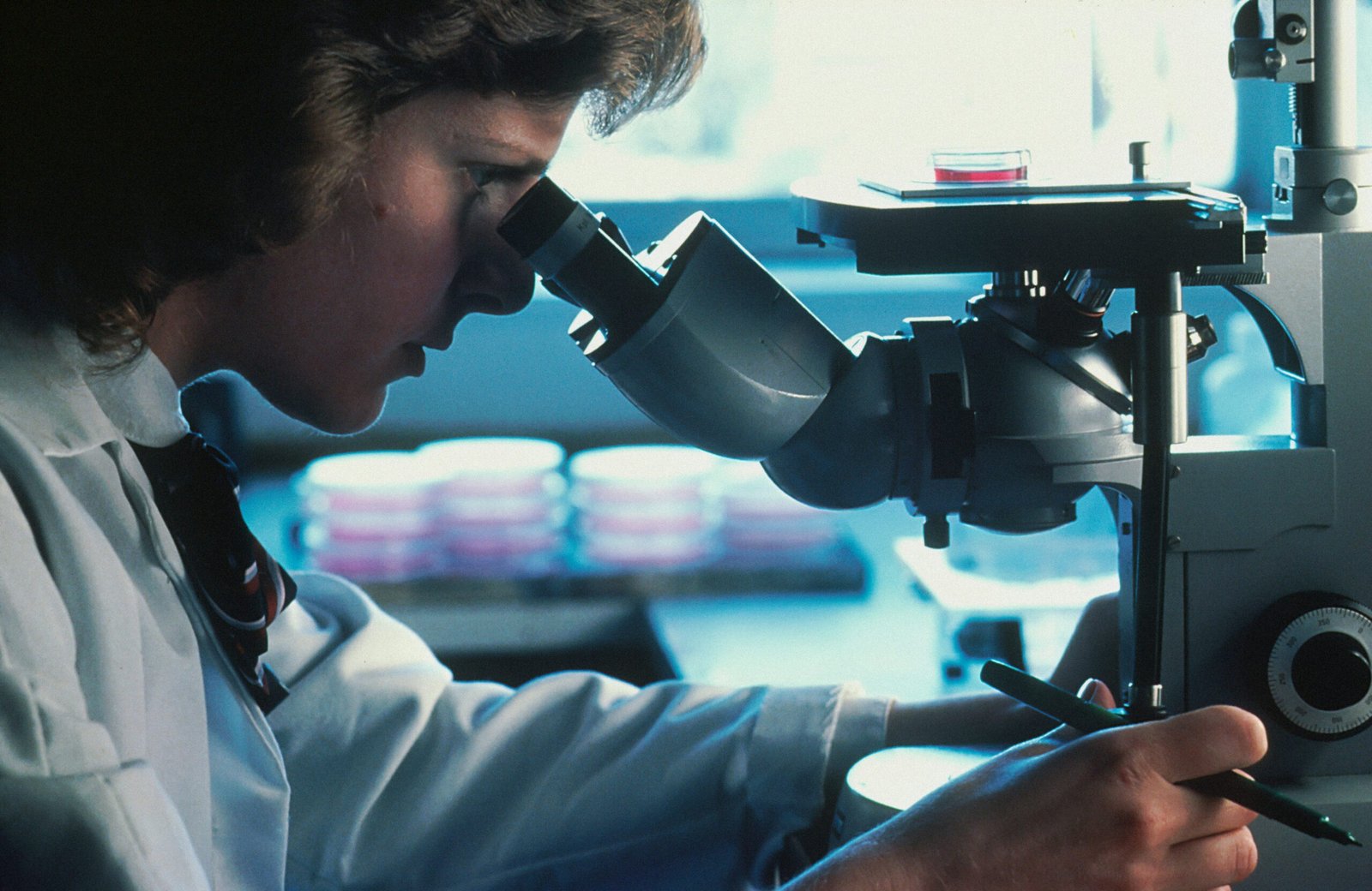

Your immune system already has cells that can find and destroy cancer. The challenge is that tumors are skilled at shutting them down. Now, a new study from McGill University has found a molecular switch that changes this balance, and the results in lab and animal studies are impressive.
Every day, immune cells called natural killer (NK) cells move through your body, searching for abnormal cells, such as infected or cancerous ones, and removing them with impressive accuracy. They do not need to learn a target in advance or require a specific antigen match. They just sense when something is wrong and act to destroy it.
This makes NK cells very appealing for cancer therapy. Unlike T-cell therapies, which require genetic matching between donor and recipient, NK cells can be expanded from umbilical cord blood or other donors and given to any patient. Scientists call this an “off-the-shelf” therapy. There is no need to wait months for custom cell manufacturing, and there is no risk of the graft attacking the patient’s own tissues.
The challenge is that solid tumors, such as glioblastoma, breast cancer, and kidney cancer, are extremely good at suppressing NK cell activity. They release molecules that weaken the immune response, disabling the very cells meant to destroy them. A landmark study in EMBO Reports by McGill University researchers has found a way to overcome this, creating NK cells that are stronger, last longer, and are much more resistant to the tumor’s defenses.
")
Two Molecular Brakes on NK Cell Power
To understand the McGill team’s work, it’s helpful to know what usually limits NK cells.
NK cells, like most immune cells, are controlled by a complex system of molecular accelerators and brakes. Two of these brakes are enzymes called PTPN1 (also known as PTP1B) and PTPN2 (also known as TC-PTP). These protein tyrosine phosphatases act as molecular switches that suppress the signals NK cells need to activate and attack tumors.
In cancer cells, studies have shown that loss of PTPN1 or PTPN2 makes tumors more susceptible to immune-mediated killing. However, it was much less clear what would happen if these same enzymes were targeted in the immune cells themselves, especially in NK cells.
The McGill researchers aimed to answer this question by using genetic tools to silence the genes for PTPN1 and PTPN2, as well as a small-molecule drug called L598 that simultaneously blocks both enzymes. They found that this approach greatly improved NK cell killing across several cancer types.
What Happens When You Release the Brakes
When PTPN1 and PTPN2 were both blocked in NK cells, either through genetic methods or with L598, several changes occurred simultaneously.
First, the NK cells became more active. They showed higher levels of the surface markers CD25 and CD69, which indicate that cells are ready to fight. The cells also grew larger, which is a known sign of NK cell activation.
Second, the cells produced much more of their cytolytic weapons, especially granzyme B and perforin, which NK cells use to penetrate cancer cell membranes and induce cell death. IFN-γ, an important immune signaling molecule, was also much higher. These are not small changes; they show the NK cells became much more aggressive at killing.
Third, and importantly, the treated NK cells killed more quickly and maintained their killing ability for longer. In experiments over time, NK cells treated with L598 killed tumor cells at almost twice the rate of untreated cells. They also kept their killing power longer because they could refill their cytolytic granules more efficiently. This supports what scientists call “serial killing,” where one NK cell destroys several tumor targets in a row.
The improved killing was seen across many cancer types, including leukemia (with four different acute myeloid leukemia lines), glioblastoma, kidney cancer, and triple-negative breast cancer, which is one of the hardest forms of the disease to treat.
The Crucial Mechanism: Amplifying the IL-2 Response
How does blocking these two phosphatases lead to these effects? The answer lies in a key signaling pathway for immune cells called JAK/STAT signaling.
When NK cells receive activation signals, particularly from a cytokine called IL-2, they transmit those signals through JAK kinases, which activate STAT transcription factors. STAT proteins then activate genes for granzyme B, perforin, IFN-γ, and other weapons. PTPN1 and PTPN2 normally act as brakes on this process, limiting NK cell responsiveness to IL-2.
When these brakes are removed, NK cells become highly sensitive to IL-2, activating STAT1, STAT3, STAT4, and STAT5 much more strongly, even at low cytokine doses. This leads to gene programs that push NK cells toward a more active, cytolytic state.
This has an important practical effect. Current NK cell therapies often require patients to get high doses of IL-2 or IL-15 from outside sources to keep the transferred immune cells alive and active. These high doses can cause serious side effects. NK cells with PTPN1/N2 inhibition might respond to lower naturally occurring cytokines, reducing the need for these toxic extra doses.
Resisting the Tumor’s Immune Defense
Beyond activating NK cells, the study addressed one of the most stubborn problems in cancer immunotherapy: the immunosuppressive tumor microenvironment.
Solid tumors do not just sit by while immune cells attack. They release molecules meant to shut down. One of the strongest of these in many cancers is a cytokine called TGFβ1 (transforming growth factor beta 1), which strongly suppresses NK cell activity by reducing their ability to kill, lowering their cytolytic weapons, and generally weakening the immune attack.
The McGill team tested what happened when NK cells were treated with PTPN1/N2 inhibitors and exposed to TGFβ1. The results showed that these enhanced cells were much more resistant. Even with TGFβ1 present, NK cells treated with L598 kept much higher levels of granzyme B than untreated cells and continued to kill tumors more effectively. This resistance seems to stem from stronger JAK/STAT signaling, which keeps NK cells active above the level at which TGFβ1 can suppress, and from AKT pathway activity that may further weaken TGFβ signaling.
This ability to resist tumor-driven immune suppression is exactly what effective solid tumor therapy needs, and it has been one of the hardest things to achieve.
From Lab to Living Models
The researchers moved beyond cell culture to test their approach in living mouse models bearing human tumors.
In mice with glioblastoma, one of the deadliest brain cancers with a median survival of about 15 months even with treatment, NK cells with dual PTPN1/N2 inhibition (either genetic or drug-based) significantly slowed tumor growth compared with untreated or standard NK cell therapy. The smallest tumors at the end of the study were always in the groups that got the enhanced NK cells.
In a separate model of triple-nIn another model using triple-negative breast cancer, mice that received NK cells pre-treated with a similar inhibitor called KQ791 had much smaller tumors compared to those given regular NK cell therapy. In this tumor model, standard NK cell therapy alone had almost no effect. KQ791 is especially interesting because it is already in Phase I clinical trials for type-2 diabetes, so it has an established safety profile and a possible path to clinical use. These effects were observed using cord blood NK cells, including cells tested against patient-derived glioblastoma cells. Cord blood is already established as a source of off-the-shelf cell therapies, with a lower risk of graft-versus-host disease and well-established logistics for banking and distribution. Demonstrating that dual inhibition enhances cord blood NK cells’ ability to kill actual patient tumor cells is a meaningful translational step.
What This Means for the Future of Cancer Treatment
There is real reason for excitement here, though it is balanced by proper scientific caution. All of the evidence so far comes from mouse models. Human clinical trials, the true test, have not yet been conducted for this specific use. The researchers also point out that there was substantial variation among cord blood donors in the extent of benefit conferred by the inhibitor, likely due to natural differences in NK cell biology. This will need to be understood before reliable clinical results can be expected.
But the combined evidence is impressive: improved killing across several cancer types, resistance to the main suppressive cytokine in the tumor microenvironment, a clear, well-understood molecular mechanism, and proven effects in animal models. With a clinically tested inhibitor compound (KQ791) that already has safety data, the path to clinical use is clearer than for many early-stage immunotherapy discoveries.
The bigger goal of this research is to create off-the-shelf NK cell therapy that does not require genetic matching, can be made in advance, stored, and administered to patients without long waits. This is one of the most exciting areas in cancer treatment. Finding that PTPN1 and PTPN2 can be adjusted as checkpoints in NK cell function adds a valuable tool to this vision.
As one of the study’s senior authors says, the research “highlights a translational opportunity to apply dual targeting of PTPN1 and PTPN2 as a strategy to improve the current NK cell-based anti-tumor immunotherapy for treating hard-to-treat cancers.” For patients with glioblastoma, triple-negative breast cancer, and other cancers with few options, this opportunity is urgently needed.
Reference:
Feng C-H, Peltier L, Chouleur T, DiPonzio M, Aubry I, Poirier AJ, et al. (2026). PTPN1/PTPN2 inhibition improves NK cancer therapy by enhancing IL-2 and mitigating TGFβ1 responses. EMBO Reports, 27: 2581–2613. https://doi.org/10.1038/s44319-026-00745-0