
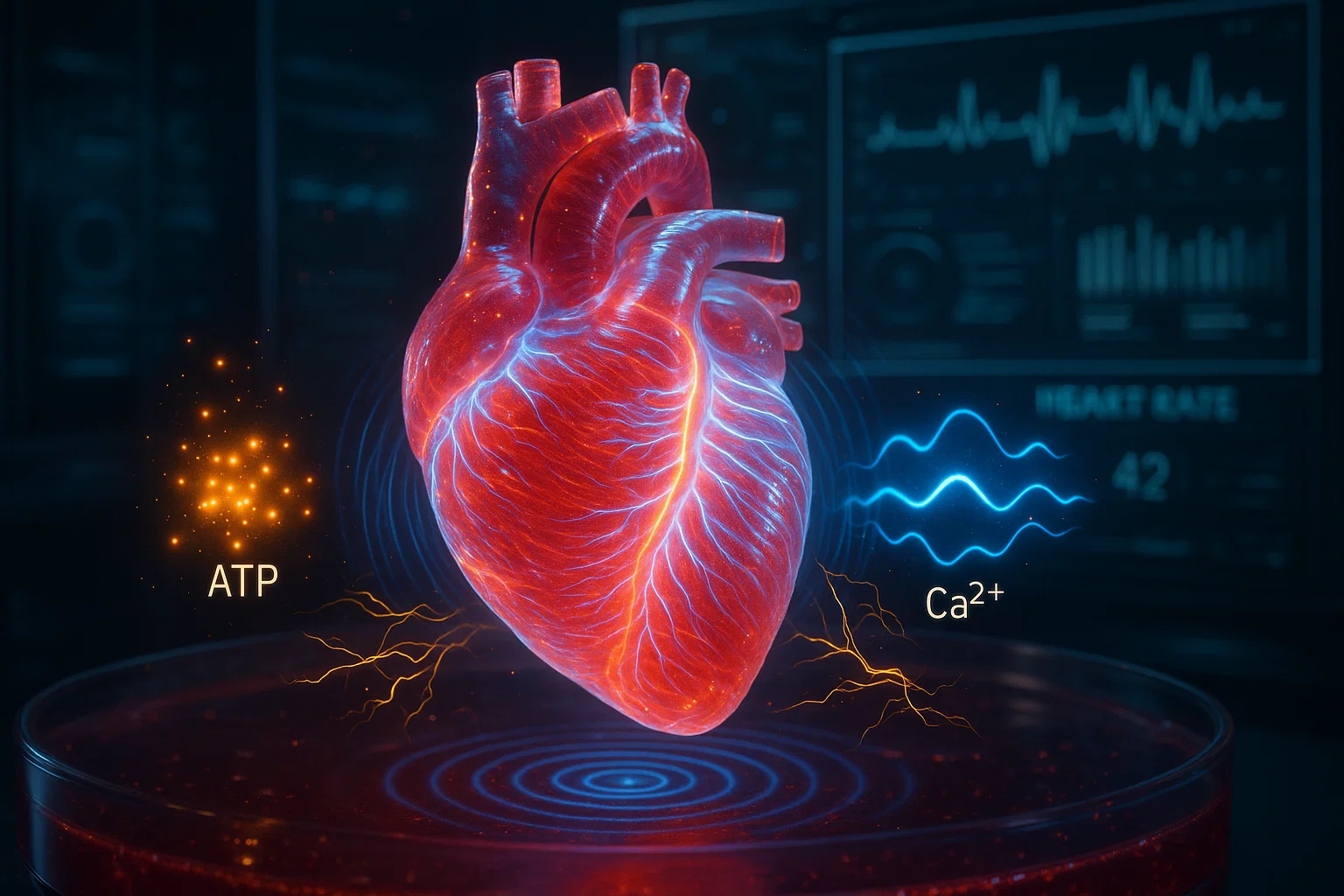

Imagine studying a human heart disease without having to take a tissue sample directly from a patient’s beating heart.
Scientists are moving closer to making this possible through an extraordinary development in biomedical engineering: human cardiac organoids, miniature three-dimensional tissues grown from stem cells that reproduce selected characteristics of the human heart.
These laboratory-grown tissues can contract, respond to medications, and provide valuable information about the molecular processes involved in cardiovascular diseases.
However, researchers have faced a persistent challenge. Cardiac cells produced from human pluripotent stem cells often resemble immature or fetal heart cells rather than fully developed adult heart muscle.
This limitation makes it difficult to study conditions that depend on the mature heart’s complex electrical, structural, and metabolic properties.
A landmark study published in Nature Cardiovascular Research in June 2025 introduced a new method for overcoming part of this challenge.
Researchers developed a directed maturation protocol that improved the structural, metabolic, and functional characteristics of human cardiac organoids.
The improved tissues successfully reproduced important features of inherited arrhythmias and desmoplakin-related cardiomyopathy. They also enabled researchers to identify a drug candidate that reduced certain disease-associated functional abnormalities in laboratory experiments.
Additional studies published in 2026 have expanded the use of cardiac organoids for investigating electrical activity, heart chamber development, and drug safety.
These findings represent important progress in cardiovascular research.
However, the organoids are experimental laboratory models. They are not fully developed miniature hearts, and the treatments identified using them have not automatically been proven effective in patients.
Why Better Heart Disease Models Are Needed
Cardiovascular diseases remain the leading cause of death worldwide.
According to the World Health Organization, an estimated 19.8 million people died from cardiovascular diseases in 2022, accounting for approximately 32% of global deaths.
These conditions include coronary artery disease, heart failure, cardiomyopathies, arrhythmias, and disorders affecting blood vessels.
Read the WHO cardiovascular disease fact sheet.
Although medical treatments have improved considerably, many cardiovascular disorders remain difficult to investigate.
One major challenge is that heart diseases often involve interactions among different cell types, genetic factors, electrical signals, metabolic processes, and structural changes.
A mutation affecting one protein may disrupt how heart muscle cells connect, leading to abnormal contraction or dangerous electrical disturbances.
Researchers therefore need experimental models that accurately reproduce these interactions.
Limitations of Traditional Research Methods
Scientists traditionally use several approaches to study cardiovascular diseases.
Animal models let researchers investigate heart function in a living organism. However, differences between human and animal biology can limit how reliably the findings translate into human treatments.
Two-dimensional cell cultures provide a relatively simple way to study human heart cells in laboratory dishes. Nevertheless, isolated cells lack much of the three-dimensional organization and mechanical environment of actual heart tissue.
Human heart tissue samples offer valuable information but are difficult to obtain and may be unavailable for studying early disease development.
These limitations have encouraged scientists to develop more sophisticated human-derived tissue models.
What Are Human Cardiac Organoids?
Human cardiac organoids, commonly abbreviated as hCOs, are three-dimensional laboratory-grown tissues that reproduce selected biological features of the human heart.
Researchers generally create them using human pluripotent stem cells.
Pluripotent stem cells can develop into many different cell types under appropriate laboratory conditions.
These include heart muscle cells, known as cardiomyocytes.
Researchers can guide stem cells through a sequence of developmental signals that encourage them to become cardiac cells.
Researchers then organize the cells into three-dimensional structures that may contain multiple interacting cell populations.
Depending on the method used, cardiac organoids can contain:
- Cardiomyocytes responsible for contraction.
- Fibroblasts involved in structural support and extracellular matrix production.
- Endothelial cells associated with blood vessels.
- Smooth muscle cells.
- Epicardial-like cells associated with the heart’s outer tissue layer.
These combinations allow scientists to study cellular interactions that are difficult to reproduce in conventional cultures.
Do Cardiac Organoids Actually Beat?
Yes. Many cardiac organoids contract spontaneously because their cardiomyocytes can generate electrical activity and coordinate mechanical contraction.
However, their beating behavior does not mean they function as complete hearts.
They typically lack the full organization, blood circulation, nervous system connections, and physiological complexity of an intact human heart.
Their value lies in reproducing particular features of cardiac biology under controlled experimental conditions.
The Major Challenge: Immature Laboratory-Grown Heart Cells
One of the biggest obstacles in cardiac organoid research is cellular immaturity.
Human pluripotent stem cell-derived cardiomyocytes often resemble developing heart cells rather than adult cardiomyocytes.
This difference affects several biological characteristics.
Structural Immaturity
Adult heart muscle cells contain highly organized contractile structures called sarcomeres.
Sarcomeres enable the coordinated contraction and relaxation of the heart.
Immature cardiomyocytes often have less developed sarcomeric organization and express different forms of structural proteins.
Metabolic Immaturity
Developing heart cells rely heavily on glucose metabolism.
As the heart matures, its metabolic processes shift toward greater mitochondrial oxidative metabolism and fatty acid utilization.
Laboratory-grown cardiomyocytes often do not fully reproduce these mature metabolic characteristics.
Electrical Immaturity
Adult heart cells have specialized ion channels and electrical properties that regulate contraction and relaxation.
Immature cardiomyocytes may beat spontaneously at different rates and respond differently to medications that influence cardiac electrical activity.
Why Does Maturation Matter?
A laboratory model that does not adequately reproduce adult heart function may miss disease mechanisms or drug responses.
For example, a medication that appears effective in immature cardiomyocytes may behave differently in mature cardiac tissue.
Improving maturation is therefore an important step toward developing more reliable preclinical models.
The Landmark 2025 Study: Maturation of Human Cardiac Organoids
In June 2025, a research team led by Mark W. Pocock and colleagues published a study titled:
Maturation of human cardiac organoids enables complex disease modeling and drug discovery.
The research appeared in Nature Cardiovascular Research, Volume 4, pages 821–840.
The authors worked across research institutions in Australia, including groups associated with the University of Queensland and the Murdoch Children’s Research Institute.
The study investigated whether researchers could reproduce biological pathways involved in normal human heart maturation.
Original Scientific Research
Read the complete study in Nature Cardiovascular Research.
The researchers developed a strategy involving two biological pathways associated with cellular energy metabolism and maturation.
These pathways involved:
AMP-activated protein kinase (AMPK) — a protein kinase that helps cells respond to changes in energy availability.
Estrogen-related receptors (ERRs) — nuclear receptors that regulate genes involved in mitochondrial metabolism and other cellular processes.
Despite their name, estrogen-related receptors are not the same as conventional estrogen receptors.
The researchers discovered that temporarily activating AMPK and ERR signaling could improve several aspects of cardiac organoid maturation.
The resulting tissues were called directed-maturation human cardiac organoids, or DM-hCOs.
How Did Scientists Make Cardiac Organoids More Mature?
The researchers used an existing experimental platform called Heart-Dyno.
Heart-Dyno is a miniaturized culture system that allows cardiac cells to organize into contracting three-dimensional tissues under mechanical loading.
The platform supports producing multiple tissue samples in a 96-well format.
This is useful because scientists can compare different biological conditions and experimental treatments.
Step 1: Producing Heart Cells From Stem Cells
Researchers began with human pluripotent stem cells.
They exposed these cells to carefully controlled developmental signals that encouraged them to differentiate into cardiac cell populations.
The resulting cells were assembled into three-dimensional heart tissue constructs.
Step 2: Encouraging Metabolic Maturation
The researchers used culture conditions designed to promote mitochondrial oxidative metabolism.
This helped move the cells toward metabolic characteristics associated with more mature heart tissue.
However, metabolic changes alone were not sufficient to reproduce all the desired adult-like properties.
Step 3: Temporarily Activating AMPK and ERR Pathways
The research team tested several compounds that influence cardiac metabolism and maturation.
Two experimental compounds were particularly important:
MK8722: An activator of AMPK signaling.
DY131: An agonist of certain estrogen-related receptors, particularly ERRβ and ERRγ.
Researchers found that a four-day treatment combining these compounds improved several indicators of cardiac maturation.
The final protocol used transient exposure rather than continuous administration.
Step 4: Evaluating Heart Tissue Function
The scientists measured properties such as contraction force, beating rate, relaxation, metabolic capacity, and gene expression.
They also compared the treated tissues with cardiac organoids produced using their earlier serum-free protocol.
The results showed that directed maturation improved several aspects of tissue function.
Importantly, the treatment was designed to mature laboratory organoids.
It was not a clinical treatment administered to patients with heart disease.
Key Discovery 1: Stronger and More Mature Heart Muscle Function
One of the clearest findings was improved contractile characteristics in the organoids.
The researchers observed increased contraction force and reduced spontaneous beating rates in several stem cell-derived organoid lines.
These changes were accompanied by increased expression of proteins associated with mature cardiac muscle.
A particularly important marker was cardiac troponin I, encoded by the TNNI3 gene.
Troponin I helps regulate contraction in heart muscle cells.
Adult heart tissue expresses a different pattern of troponin isoforms from immature cardiac tissue.
The increased expression of mature cardiac troponin I therefore provided evidence that the organoids had progressed toward a more developed functional state.
Researchers also examined other sarcomeric proteins involved in cardiac contraction.
These measurements supported the conclusion that the directed-maturation protocol improved the contractile machinery of the tissues.
Why Is This Important?
A more mature contractile system can make laboratory heart models more useful for studying diseases associated with impaired heart muscle function.
However, the findings do not mean that the organoids became identical to adult human heart tissue.
They displayed more adult-like properties in selected measurements while retaining limitations characteristic of laboratory-grown models.
Key Discovery 2: Improved Mitochondrial Metabolism
The heart requires substantial energy to contract continuously.
Mitochondria produce much of this energy through oxidative phosphorylation.
During normal cardiac development, cardiomyocytes’ metabolic characteristics change considerably.
The researchers found that the directed-maturation protocol increased the expression of oxidative phosphorylation proteins.
The treated cardiac organoids also demonstrated increased metabolic capacity.
Researchers used experimental techniques to investigate mitochondrial respiration and cells’ ability to respond to changing energy demands.
These measurements showed that directed-maturation organoids had greater maximal respiratory capacity than the comparison tissues.
What Does This Mean?
The results suggest that the treated organoids developed metabolic characteristics more closely resembling mature heart muscle.
This is significant because mitochondrial function contributes to several cardiovascular diseases.
However, increased metabolic capacity in a laboratory model does not necessarily mean that the tissue can reproduce every aspect of adult cardiac metabolism.
The findings represent an improvement in experimental modeling rather than complete biological maturation.
Key Discovery 3: The Organoids Contained Multiple Human Heart Cell Types
Cardiovascular diseases rarely involve cardiomyocytes alone.
Heart muscle cells interact with fibroblasts, vascular cells, and other supporting populations.
These interactions influence structural organization, electrical activity, and disease progression.
The researchers used single-nucleus RNA sequencing to examine the cellular composition of their cardiac organoids.
This technique measures gene expression in individual cell nuclei.
The analysis identified several cell populations resembling those found in native human heart tissue.
These included cardiomyocytes, fibroblasts, endothelial cells, smooth muscle cells, and epicardial-like cells.
Why Does Cellular Complexity Matter?
Consider cardiac fibrosis.
Fibrosis occurs when excess extracellular matrix accumulates in heart tissue, often contributing to stiffness and impaired function.
Fibroblasts are central to this process.
A model containing only cardiomyocytes may fail to reproduce important interactions between damaged heart muscle and fibrotic tissue.
Multicellular cardiac organoids can provide additional information about these processes.
Nevertheless, the study also identified important differences between organoids and the human heart.
The organoids contained a higher proportion of cardiomyocytes, fewer endothelial cells, and lacked some populations, including immune and neural cells.
They therefore reproduced selected aspects of cardiac cellular complexity rather than the full tissue environment.
Key Discovery 4: Modeling Inherited Heart Rhythm Disorders
A particularly valuable application of the new organoids was studying inherited arrhythmias.
Arrhythmias occur when the electrical signals controlling heartbeat become abnormal.
Some can cause palpitations or dizziness, while certain severe arrhythmias may lead to cardiac arrest.
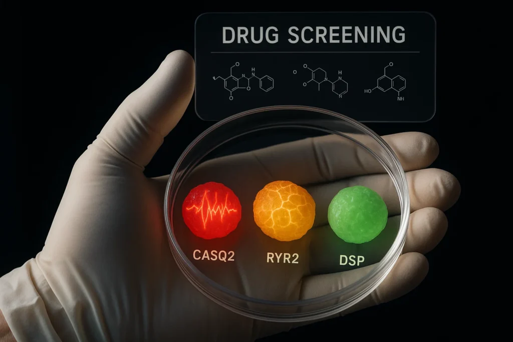
The researchers investigated organoids containing mutations in two genes:
CASQ2, Calsequestrin 2
This gene encodes a protein that stores and regulates calcium within heart muscle cells.
RYR2, Ryanodine receptor 2
This gene encodes a calcium-release channel that helps coordinate cardiac contraction.
Mutations in these genes can cause inherited disorders such as catecholaminergic polymorphic ventricular tachycardia (CPVT).
CPVT is associated with dangerous stress- or exercise-triggered ventricular arrhythmias.
What Did the Organoids Reveal?
When researchers produced mature cardiac organoids from stem cells carrying CASQ2 or RYR2 mutations, the tissues showed characteristics linked to increased susceptibility to arrhythmias.
This included abnormal responses related to calcium handling and electrical stability.
These findings suggested that the improved organoids could reproduce certain disease-related functional changes more effectively than less mature models.
Why Is This a Major Advance?
Inherited arrhythmia disorders can be difficult to study using isolated cells because the surrounding tissue environment influences electrical behavior.
Three-dimensional organoids provide an opportunity to examine coordinated contraction and disease-associated abnormalities.
However, laboratory pro-arrhythmic behavior does not reproduce every clinical feature of CPVT.
The findings support the model’s usefulness for research, not its direct use as a diagnostic test.
Key Discovery 5: Modeling Desmoplakin-Related Cardiomyopathy
Another major achievement involved a condition associated with mutations in the DSP gene.
DSP encodes desmoplakin, a structural protein that helps connect neighboring heart muscle cells through specialized cellular junctions.
These connections are important because heart tissue experiences continuous mechanical stress during contraction.
Certain DSP mutations are associated with inherited cardiomyopathies that may involve:
- Impaired cardiac contraction.
- Abnormal tissue organization.
- Myocardial fibrosis.
- Increased risk of ventricular arrhythmias.
- Heart failure in some patients.
The researchers produced cardiac organoids carrying a disease-associated DSP mutation.
They then compared their behavior with genetically corrected control organoids.
What Did the Scientists Observe?
The mutant organoids developed important disease-like characteristics.
These included increased fibrotic changes and impaired contractile function.
The researchers also identified molecular patterns associated with extracellular matrix remodeling and cellular stress.
Importantly, some of these features resembled changes observed in human heart tissue associated with DSP-related cardiomyopathy.
This provided evidence that the organoids could reproduce aspects of a complex inherited heart disease involving more than one cellular pathway.
Key Discovery 6: Identifying an Experimental Drug Candidate
The researchers next investigated whether the disease model could help identify compounds that improve abnormal cardiac function.
They evaluated several experimental treatments using the DSP-mutant organoids.
One compound, INCB054329, produced a promising response.
INCB054329 is a bromodomain and extra-terminal domain (BET) inhibitor.
BET proteins regulate gene expression.
Researchers have investigated BET inhibition in several disease areas because these proteins influence biological pathways associated with inflammation, cellular growth, and tissue remodeling.
What Happened After Treatment?
In the laboratory experiments, INCB054329 mitigated certain functional defects associated with the DSP mutation.
The researchers also observed changes in the expression of some disease-associated proteins.
However, the compound did not fully reverse the entire molecular disease signature.
Several extracellular matrix-associated proteins remained elevated.
This is an important distinction.
The experiment showed that the organoid model could identify a compound that modifies specific disease-related responses. It did not demonstrate a cure for desmoplakin-related cardiomyopathy.
INCB054329 has not been approved a treat this inherited heart condition.
Further research is needed to assess its safety, effectiveness, and potential clinical relevance.
Why Are Mature Cardiac Organoids Valuable for Drug Discovery?
Developing a new cardiovascular medicine is a lengthy and complex process.
A candidate drug may appear promising in simple laboratory experiments but fail when tested in more realistic biological systems.
One reason is that isolated cells cannot fully reproduce how tissues respond to medication.
Cardiac organoids may help address some of these limitations.
Testing Drug Effects on Heart Contraction
Researchers can measure whether a drug increases or decreases contraction force.
They can also investigate changes in beating rate and relaxation.
These measurements show how a candidate compound affects cardiac mechanical function.
Investigating Electrical Side Effects
Some medications can alter cardiac ion channels and potentially increase arrhythmia risk.
Cardiac organoids may help researchers identify abnormal electrical responses during preclinical testing.
Studying Inherited Disease Mechanisms
Organoids carrying specific genetic variants can help scientists investigate how mutations influence tissue function.
This approach may be useful when studying rare inherited cardiovascular disorders.
Comparing Experimental Treatments
The Heart-Dyno platform supports multiple cardiac organoids in parallel.
This may allow researchers to compare different compounds and concentrations more efficiently than some traditional tissue systems.
However, improved laboratory screening does not eliminate the need for animal studies where appropriate, human clinical trials, or regulatory safety evaluations.
New Research in 2026: Cardiac Organoids Become More Sophisticated
Since the original 2025 study, researchers have continued developing cardiac organoid technology.
Several 2026 publications demonstrate the growing range of applications.
1. Chamber-Like Cardiac Organoids June 2026
A study published in Nature Communications on June 2, 2026, described self-assembled cardiac organoids that reproduce selected structural characteristics of developing heart chambers.
The paper was titled:
Self-assembled chamber-like cardiac organoids for modeling cardiac chamber formation and cardiotoxicity assessment.
Read the study in Nature Communications.
The researchers generated structures containing myocardial tissue, endothelial-like layers, and an internal chamber.
These organoids displayed spontaneous contraction and calcium signaling.
Their architecture offered new opportunities to investigate early heart development and drug-related cardiac toxicity.
The research is particularly relevant because conventional cardiac organoids often lack the chamber-like organization of the developing heart.
However, these structures primarily model aspects of cardiac development rather than the complete anatomy and function of an adult heart.
2. Drug Toxicity and Disease Modeling June 2026
Another study, published in Stem Cell Research & Therapy on June 17, 2026, investigated the use of induced pluripotent stem cell-derived cardiac organoids for drug testing.
The paper was titled:
iPSC-derived cardiac organoids for drug cardiotoxicity evaluation and efficacy prediction in myocardial infarction and cardiac hypertrophy models.
The research explored how cardiac organoids could support laboratory evaluation of drug toxicity and treatment responses in experimental models of heart injury and hypertrophy.
This work reflects the growing interest in using human-derived tissue models for more disease-relevant preclinical testing.
Nevertheless, identifying a drug response in an experimental organoid does not establish that the same treatment will improve clinical outcomes.
In this research context, efficacy prediction should not be confused with a validated ability to predict treatment effectiveness in individual patients.
3. Advanced Electrical Testing September 2026
A study published in Pharmacological Research in September 2026 examined how cardiac organoids could be used to investigate electrical activity and screen potential antiarrhythmic compounds.
The paper was titled:
Modeling cardiac electrical activity using cardiac organoids for drug screening.
Researchers combined several experimental methods, including:
- Multielectrode array measurements.
- Optical mapping of calcium signals.
- Patch-clamp electrophysiology.
These techniques let researchers examine electrical activity in the organoids at different levels.
The study investigated responses to compounds including E-4031, cisapride, and ATX-II.
The researchers reported that some electrical and calcium-related responses in cardiac organoids more closely resembled cardiac tissue than conventional two-dimensional cardiomyocyte cultures.
These results support the potential use of cardiac organoids in preclinical arrhythmia research.
However, further validation is required before such platforms can reliably predict drug-associated arrhythmia risk in humans.
How Do Cardiac Organoids Compare With Other Heart Research Models?
Different laboratory and animal models provide different types of information.
| Research approach | Main strength | Important limitation |
| 2D cardiomyocyte cultures | Relatively accessible and useful for molecular experiments | Limited tissue organization |
| Animal models | Support whole-organism research | Species differences may affect translation |
| Engineered cardiac tissues | Allow mechanical and functional measurements | May lack full cellular complexity |
| Human cardiac organoids | Combine human-derived cells with three-dimensional organization | Often remain developmentally immature |
| Directed-maturation cardiac organoids | Improved selected structural, metabolic, and functional characteristics | Do not fully reproduce adult human heart physiology |
| Human clinical studies | Directly assess outcomes in people | Require careful ethical oversight, substantial resources, and time |
No single experimental approach is sufficient for every research question.
The most reliable findings often emerge when researchers combine complementary models.
Could Cardiac Organoids Support Personalized Medicine?
One long-term possibility is developing disease models from a patient’s own cells.
Scientists can produce induced pluripotent stem cells from certain adult cells and then guide them to become cardiomyocytes.
If those cells carry a disease-associated genetic variant, the resulting cardiac tissue may reproduce some effects of that mutation.
Researchers could potentially use these models to compare experimental treatments.
For example, cardiac organoids derived from a person with an inherited cardiomyopathy might reveal how a particular mutation affects contractile function.
This information could help scientists understand disease mechanisms and investigate possible therapeutic targets.
However, the concept has important limitations.
Different stem cell lines may behave differently.
Organoid manufacturing processes can also introduce variability.
Furthermore, a drug that improves tissue function in an organoid may not provide the same benefit in a living person.
Patient-specific cardiac organoid testing is not yet a established clinical method for selecting cardiovascular medications.
Prospective validation would be required before such testing could routinely guide treatment decisions.
What Are the Main Limitations of Mature Cardiac Organoids?
Although the technology has progressed, several scientific challenges remain.
1. Incomplete Adult-Like Maturation
The 2025 study improved several maturity indicators, but the organoids did not become identical to fully developed adult heart tissue.
Some important structural and physiological characteristics remained different.
2. Limited Cellular Diversity
The models contained multiple cardiac cell types but lacked certain populations, including immune and neural cells.
These missing components may influence disease development and treatment response.
3. Lack of Complete Organ-Level Physiology
Cardiac organoids cannot fully reproduce the interactions among heart chambers, coronary circulation, nervous system regulation, and whole-body metabolism.
4. Differences Between Stem Cell Lines
Genetic background and cell manufacturing methods can influence how organoids develop.
Reproducibility across laboratories and patient-derived cell lines remains a key concern.
5. Limited Ability to Model Long-Term Disease
Many cardiovascular conditions develop over years or decades.
Organoid experiments often occur over much shorter periods.
This makes it difficult to reproduce every stage of chronic disease progression.
6. Clinical Drug Responses Remain Uncertain
A promising response in an organoid helps generate hypotheses and select compounds for further study.
It does not establish that the same compound will be safe or effective in patients.
These limitations explain why cardiac organoids are best understood as improving preclinical research rather than replacing clinical medicine.
What Could the Future of Cardiac Organoid Research Look Like?
Researchers are investigating several directions that may improve the usefulness of cardiac organoids.
More Complete Tissue Maturation
Future approaches may combine metabolic stimulation, mechanical loading, electrical pacing, and carefully controlled signaling pathways.
The goal is to reproduce additional adult-like cardiac characteristics.
Improved Vascular Organization
Developing more sophisticated blood vessel-like networks could help researchers investigate oxygen delivery, nutrient transport, and interactions between cardiac muscle and vascular cells.
Integration of Immune and Neural Cells
Including more cell populations may allow scientists to investigate inflammatory responses and nervous system influences on heart function.
Higher-Throughput Drug Screening
Automated systems could eventually allow researchers to test larger collections of compounds while measuring contraction, electrical activity, and cellular stress.
Better Modeling of Rare Genetic Diseases
Stem cell-derived cardiac tissues may provide new opportunities to study disorders for which human heart samples are difficult to obtain.
However, each proposed application will require its own scientific validation.
What Does This Research Mean for Heart Disease Patients?
The research offers an important reason for optimism about the future of cardiovascular drug development.
More realistic human-derived models may help scientists identify disease mechanisms that are difficult to study using conventional methods.
They may also support earlier detection of potentially harmful drug effects and provide a better understanding of inherited heart disorders.
However, this technology has not yet produced a proven cure for cardiomyopathy, arrhythmia, or heart failure.
Patients should not interpret the laboratory findings as evidence that treatment based on cardiac organoids is currently available.
People with suspected or diagnosed heart disease should continue to rely on established medical evaluation and treatments recommended by qualified healthcare professionals.
For information about cardiovascular disease prevention and management, see the World Health Organization’s cardiovascular disease resource.
Conclusion: A Major Advance in Human Heart Disease Modeling
The development of more mature human cardiac organoids marks an important step in cardiovascular research.
The 2025 study by Pocock and colleagues demonstrated that temporary activation of AMPK and estrogen-related receptor pathways could improve several aspects of laboratory-grown heart tissue.
These improvements included more mature sarcomeric proteins, stronger contraction, increased metabolic capacity, and more representative responses to selected cardiac medications.
The researchers also demonstrated that the organoids could reproduce features of inherited arrhythmias and desmoplakin-related cardiomyopathy.
Importantly, the model helped identify an experimental compound that reduced certain functional abnormalities associated with a DSP mutation.
Research published in 2026 has continued expanding the field through new organoid structures, electrical testing techniques, and drug-screening applications.
Together, these findings suggest that cardiac organoids could become increasingly useful for investigating cardiovascular diseases and evaluating potential treatments.
However, significant work remains before the technology can routinely guide clinical decisions.
The central breakthrough is not that scientists have created fully functional replacement hearts. It is that they have developed more sophisticated laboratory models that reproduce important aspects of human heart biology and disease.
With continued improvement and validation, these models may help researchers understand cardiovascular disorders more precisely and develop safer, more effective treatments.
References
1. Pocock MW, Reid JD, Robinson HR, et al. (2025).
Maturation of human cardiac organoids enables complex disease modeling and drug discovery.
Nature Cardiovascular Research, 4, 821–840.
Published June 25, 2025.
https://doi.org/10.1038/s44161-025-00669-3
PubMed: https://pubmed.ncbi.nlm.nih.gov/40562874/
2. Zou X, et al. (2026).
Self-assembled chamber-like cardiac organoids for modeling cardiac chamber formation and cardiotoxicity assessment.
Nature Communications, 17, Article 7096.
Published June 2, 2026.
https://doi.org/10.1038/s41467-026-73822-6
3. Hou C, Zhang Q, Zhang Z, et al. (2026).
iPSC-derived cardiac organoids for drug cardiotoxicity evaluation and efficacy prediction in myocardial infarction and cardiac hypertrophy models.
Stem Cell Research & Therapy, 17, Article 296.
Published June 17, 2026.
https://doi.org/10.1186/s13287-026-05079-1
4. Zhang Q, Yang N, Chen J, et al. (2026).
Modeling cardiac electrical activity using cardiac organoids for drug screening.
Pharmacological Research, 231, 108373.
Published September 2026.
https://doi.org/10.1016/j.phrs.2026.108373
PubMed: https://pubmed.ncbi.nlm.nih.gov/42567459/
5. World Health Organization. (2025).
Cardiovascular diseases (CVDs).
https://www.who.int/news-room/fact-sheets/detail/cardiovascular-diseases-(CVDs)