

For decades, electronic pacemakers have helped millions of people live with dangerously slow or irregular heart rhythms. These devices deliver carefully timed electrical impulses that keep the heart beating when its natural electrical system cannot function properly.
But conventional pacemakers come with an engineering challenge: they typically require a generator implanted beneath the skin and electrical wires, called leads, that connect the generator to the heart.
These components can sometimes cause complications, including infections, lead fractures, and problems involving blood vessels or heart valves.
Scientists and medical-device manufacturers are now exploring two distinct approaches to improving cardiac pacing.
The first involves leadless pacemakers, miniature devices implanted directly inside the heart without traditional transvenous leads or a chest generator pocket.
The second involves biological pacemakers, experimental therapies that attempt to create natural pacemaker-like activity by modifying living heart cells.
In recent years, leadless devices such as Medtronic’s Micra and Abbott’s Aveir have expanded the treatment options available to patients requiring permanent pacing.
Meanwhile, researchers at Cedars-Sinai have made progress using gene therapy to reprogram cardiac cells into cells capable of generating their own electrical impulses.
Research published in 2026 provides further evidence of the potential of both approaches.
However, an important distinction remains: leadless electronic pacemakers are established clinical technologies for selected patients, while biological pacemakers are still being investigated in preclinical research.
Understanding their differences helps explain where cardiac rhythm management is heading.
What Is a Pacemaker, and Why Do Some People Need One?
The human heart has its own electrical conduction system.
Under normal circumstances, each heartbeat begins in a small group of specialized cells called the sinoatrial node, or SA node, located in the right atrium.
Often described as the heart’s natural pacemaker, the SA node generates electrical impulses that spread through the atria.
These impulses then reach the atrioventricular (AV) node and travel through specialized conduction pathways to activate the ventricles, which pump blood to the lungs and the rest of the body.
When this system fails, the heart may beat too slowly or become poorly coordinated.
Bradycardia: When the Heart Beats Too Slowly
Bradycardia generally refers to a resting heart rate below 60 beats per minute.
However, a slow heart rate does not always indicate disease. Healthy athletes and people who are sleeping may have low heart rates without experiencing problems.
Pacemaker treatment may be necessary when abnormal electrical conduction produces symptoms or clinically important risks.
Possible symptoms include:
- Recurrent dizziness.
- Fainting or near-fainting.
- Unusual fatigue.
- Exercise intolerance.
- Shortness of breath.
- Weakness associated with an inadequate heart rate.
Common conditions that may require pacing include sinus node dysfunction and certain forms of atrioventricular block.
Pacemakers can also be used in other specific clinical circumstances.
Treatment decisions depend on the underlying condition, symptoms, medical history, and professional assessment rather than heart rate alone.
How Do Traditional Pacemakers Work?
A conventional transvenous pacemaker usually contains a pulse generator implanted under the skin in the upper chest.
One or more insulated electrical leads pass through a vein into the heart.
The generator monitors the heart’s electrical activity and delivers pulses when pacing is needed.
Modern systems may adjust pacing rates according to activity and support coordination between different heart chambers.
Traditional pacemakers have a long history of effectiveness and remain appropriate for many patients.
However, their leads and generator pockets create potential sources of complications.
What Problems Can Traditional Pacemakers Cause?
Some patients experience lead displacement, insulation failure, conductor fracture, or complications involving the veins used to introduce the leads.
Others may develop infections involving the generator pocket or the implanted hardware.
Leads crossing the tricuspid valve can also contribute to valve dysfunction in some circumstances.
These risks are clinically important, but their frequency depends on patient characteristics, device type, implantation technique, and duration of follow-up.
It would be misleading to claim that a fixed percentage of all long-term pacemaker complications occurs because of leads or generator pockets without identifying the supporting study and its definitions.
The goal of leadless pacing is to eliminate certain hardware-related risks while maintaining effective electrical stimulation.
What Are Leadless Pacemakers?
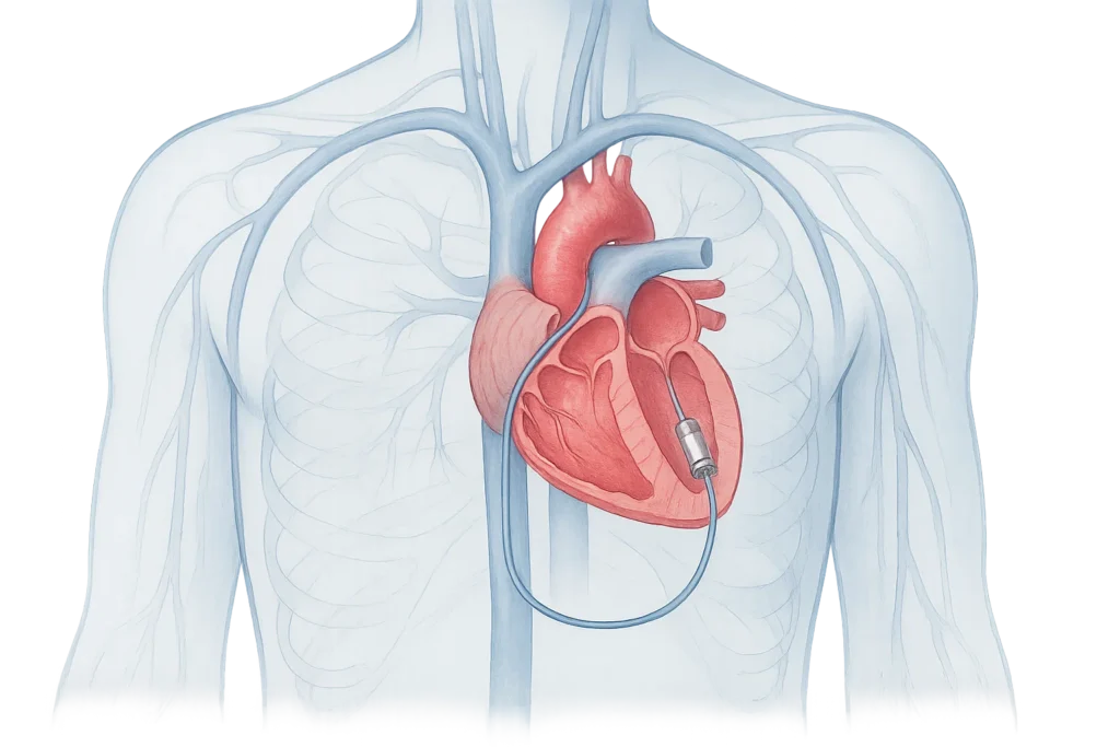
Leadless pacemakers are miniature self-contained electronic devices implanted directly into a heart chamber.
Unlike traditional pacemakers, they generally do not require a separate generator pocket beneath the skin or long transvenous leads connecting the chest to the heart.
The device contains its own battery, electronic circuitry, sensing components, and pacing electrodes.
How Are Leadless Pacemakers Implanted?
A cardiologist specializing in heart rhythm disorders typically inserts a delivery catheter through a large vein, often in the groin.
The catheter is guided into the heart using imaging.
The miniature pacemaker is then positioned and attached to the inner heart wall.
Once its electrical performance and fixation are verified, the delivery system is removed.
This method avoids the chest incision and generator pocket required for a conventional transvenous pacemaker.
However, it is still an invasive cardiac procedure.
Potential complications include bleeding, vascular injury, device dislodgement, infection, cardiac perforation, and pericardial effusion.
Leadless does not mean risk-free.
Medtronic Micra: A Miniaturized Pacemaker Inside the Heart
Medtronic’s Micra family helped establish leadless pacing as a clinical treatment option.
The first Micra system received US Food and Drug Administration approval in 2016.
Later generations introduced updated programming capabilities and improved projected battery longevity.
In April 2023, the FDA approved two newer models:
- Micra VR2, designed for ventricular pacing.
- Micra AV2, which uses mechanical sensing to help coordinate ventricular pacing with atrial contraction in selected patients.
Micra devices are implanted inside the right ventricle.
Their small size eliminates the need for traditional chest-pocket implantation.
How Does Micra AV2 Maintain Coordination?
Micra AV2 uses an accelerometer to detect mechanical activity associated with atrial contraction.
This information allows the device to time certain ventricular pacing impulses in relation to atrial activity.
The technique can improve atrioventricular coordination in appropriately selected patients.
However, it is important to understand that Micra AV2 does not directly pace the atrium.
It is a ventricular leadless pacemaker that uses mechanical sensing to support atrioventricular synchrony.
FDA source: Micra AV2 and VR2 Approval Record
Manufacturer announcement: Medtronic, FDA Approval of Next-Generation Micra Systems
Abbott Aveir: Expanding Leadless Pacing to Two Heart Chambers
Abbott’s Aveir technology represents another major development.
The Aveir VR leadless pacemaker received FDA approval in March 2022.
In June 2023, the FDA approved the Aveir DR system, introducing a dual-chamber leadless pacing approach.
Unlike a single ventricular leadless pacemaker, Aveir DR uses two separate implanted devices:
- An atrial pacemaker positioned in the right atrium.
- A ventricular pacemaker positioned in the right ventricle.
The two devices communicate with one another to coordinate pacing activity.
This is a significant development because it allows both atrial and ventricular pacing without conventional long leads.
How Do Two Leadless Pacemakers Communicate?
Aveir DR uses proprietary implant-to-implant communication technology.
The devices exchange information through conductive communication signals to coordinate electrical stimulation.
This supports atrioventricular synchrony, helping the atria and ventricles contract in the appropriate sequence.
The system therefore expands the range of patients who may be considered for leadless pacing.
FDA approval record: Aveir DR Leadless System
What Did the Clinical Trials Reveal?
A pivotal clinical study of the dual-chamber Aveir system was published in The New England Journal of Medicine in 2023.
The study enrolled 300 patients who had established indications for dual-chamber pacing.
Among them, 190 had sinus node dysfunction and 100 had atrioventricular block as their primary pacing indication.
The researchers examined implantation success, safety, atrial electrical performance, and coordination between the atria and ventricles.
Key Results
The procedure successfully implanted two functioning leadless pacemakers with communication between them in 295 of 300 patients, equivalent to 98.3%.
The study found that 90.3% of patients were free from device- or procedure-related serious adverse events through 90 days.
Adequate atrial pacing threshold and sensing performance were achieved in 90.2% of patients.
The study also reported that 97.3% of patients achieved its predefined atrioventricular synchrony performance endpoint.
These findings supported the feasibility of a dual-chamber leadless system.
However, the study was not a randomized comparison against conventional dual-chamber pacemakers, and its main published outcomes focused on relatively early follow-up.
Original clinical trial: Knops et al., A Dual-Chamber Leadless Pacemaker, NEJM (2023)
Are Leadless Pacemakers Safer Than Traditional Pacemakers?
Leadless pacemakers avoid some of the hardware-related problems associated with conventional devices.
However, overall safety depends on the type of device, the clinical indication, and the individual patient.
A leadless system may reduce certain complications while introducing different procedural challenges.
This means safety must be evaluated using carefully designed comparisons.
New Evidence From 2026
A study published in 2026 compared early real-world outcomes of dual-chamber Aveir leadless pacemakers with conventional transvenous dual-chamber systems using US Medicare data.
The analysis included:
- 759 patients receiving dual-chamber leadless pacemakers.
- 77,422 patients receiving transvenous dual-chamber pacemakers.
After adjustment for measured differences between patient groups, device-related complications at six months occurred in 2.8% of the leadless group and 5.9% of the transvenous group.
Device reinterventions were also less frequent in the leadless group, occurring in 2.1% versus 4.3%.
Overall adjusted complications at six months were 4.1% versus 6.9%.
However, the study did not find a statistically significant difference in mortality.
Because this was observational research, residual confounding and differences in clinical selection remain possible.
The results support the potential safety benefits of leadless pacing, but they do not establish that leadless systems are the best option for every patient.
2026 clinical research: Leadless Versus Transvenous Dual-Chamber Pacemakers, PubMed
What Are the Remaining Limitations of Leadless Pacemakers?
Despite their advantages, leadless devices have several limitations.
Battery Replacement and Device Management
Leadless pacemakers contain batteries with finite lifespans.
When a device reaches the end of its useful life, physicians must decide whether it should be retrieved, deactivated and left in place, or managed through another pacing strategy.
The appropriate decision depends on the device, implantation duration, surrounding tissue, and available space within the heart.
Device Retrieval
Some leadless pacemakers are designed with retrieval features.
However, removing a device after years of implantation may be more complicated because tissue can grow around it.
Long-term management remains an important area of research.
Implantation Risks
Although leadless devices avoid chest pockets and traditional leads, their implantation can still cause serious complications.
Cardiac perforation, pericardial effusion, vascular injury, and device dislodgement are among the potential concerns.
Limited Pacing Applications
Not every leadless system provides the same functions as a traditional pacemaker.
Patients requiring particular pacing configurations, cardiac resynchronization therapy, defibrillation, or specialized conduction-system pacing may need other devices or approaches.
Cost and Availability
Leadless systems may have higher initial device costs and require specialized expertise.
Availability and insurance coverage vary across healthcare systems and countries.
What Are Biological Pacemakers?
While leadless pacemakers improve the design of electronic cardiac devices, biological pacemakers take a fundamentally different approach.
Instead of implanting an electronic device that generates pacing pulses, researchers attempt to create living cells capable of producing pacemaker-like electrical activity.
The long-term goal is to restore some of the heart’s natural electrical function.
How Does the Heart Generate Its Own Rhythm?
The sinoatrial node contains specialized cells that spontaneously generate electrical impulses.
This property is known as automaticity.
The cells contain ion channels and molecular machinery that allow their electrical potential to change rhythmically without requiring a signal from another part of the heart.
Their activity also responds to signals from the autonomic nervous system.
For example, heart rate usually increases during exercise and decreases during rest.
A biological pacemaker would ideally reproduce these important characteristics.
Gene Therapy: Reprogramming Heart Cells Into Pacemaker-Like Cells
One of the most extensively investigated approaches involves a transcription factor called TBX18.
A transcription factor is a protein that influences the activity of many genes.
TBX18 plays an important role during cardiac development.
Researchers have investigated whether introducing TBX18 into ordinary heart muscle cells can change their molecular identity and electrical behavior.
The goal is to convert some existing cardiomyocytes into cells with characteristics resembling natural pacemaker cells.
Why Is TBX18 Important?
Ordinary working heart muscle cells generally require an electrical impulse from another cell to initiate contraction.
Pacemaker cells, by contrast, can initiate their own rhythmic electrical activity.
In experimental studies, TBX18 gene delivery has altered gene expression and increased spontaneous electrical activity in targeted cardiac tissue.
Researchers often use modified viral vectors to transport genetic instructions into cells.
These vectors are engineered for research or therapeutic delivery, but their safety and biological effects still require careful evaluation.
A biological pacemaker therefore does not necessarily involve transplanting new cells.
It may instead involve reprogramming cells already present in the heart.
January 2026 Breakthrough: AAV-TBX18 Biological Pacing
An important study from Cedars-Sinai was published in the January 2026 issue of Molecular Therapy.
The paper, titled Biological Pacemaker Induced by Focal Cardiac Transduction With AAV-TBX18, investigated whether a modified adeno-associated virus could deliver TBX18 and generate pacemaker-like activity.
Earlier experiments had used adenoviral delivery, but immune responses could limit how long the modified cells remained active.
The research team therefore investigated an adeno-associated viral vector, known as AAV-TBX18.
What Did Researchers Find?
In rats, targeted AAV-TBX18 delivery changed local cardiac gene expression toward a profile resembling the sinoatrial node.
It also produced spontaneous electrical activity at the injection site.
The induced pacemaker activity showed responsiveness to autonomic influences and was associated with improved exercise tolerance.
The investigators also tested catheter-based AAV-TBX18 delivery in pigs with experimentally induced complete atrioventricular block.
The treated pigs showed increased heart rates compared with control animals.
The effect remained evident for at least six weeks in rats and four weeks in pigs during the reported observation periods.
These findings demonstrated that targeted gene delivery could produce physiologically responsive pacing activity in animal models.
However, the study did not demonstrate that the treatment could safely replace a permanent electronic pacemaker in humans.
Original research: Molecular Therapy (2026), DOI: 10.1016/j.ymthe.2025.10.037
PubMed record: Biological Pacemaker Induced by Focal Cardiac Transduction With AAV-TBX18
August 2026 Breakthrough: Restoring Pacemaker Activity in Pigs With Sinus Node Dysfunction
A second important study was published online in JACC: Clinical Electrophysiology in August 2026.
Researchers led by James Dawkins and colleagues at Cedars-Sinai investigated whether TBX18 gene delivery could create a functional biological pacemaker in pigs with experimentally induced sick sinus syndrome.
How Was the Experiment Conducted?
The researchers created a large-animal model of severe sinus node dysfunction.
They used targeted radiofrequency ablation to disrupt the natural sinoatrial node.
Backup electronic pacemakers were implanted to support the animals when necessary.
After confirming the development of sinus node dysfunction, the investigators delivered either a TBX18-containing adenoviral vector or a control solution into the lower atrial septum.
Nine pigs received TBX18 treatment, and nine received the control intervention.
Researchers monitored cardiac rhythm for four weeks after injection.
What Were the Results?
Two weeks after treatment, pigs receiving TBX18 demonstrated significantly higher intrinsic heart rates than controls.
The researchers reported average heart rates of approximately:
- 96.3 beats per minute in TBX18-treated animals.
- 71.4 beats per minute in control animals.
The treated animals also relied less on their backup electronic pacemakers.
Analysis of heart rate variability suggested improved rhythm stability, with fewer pauses and certain abnormal rhythm episodes.
Additional cellular testing identified spontaneous electrical activity and characteristics resembling sinoatrial node cells near the injection site.
These results support the concept that targeted gene therapy can convert some heart muscle cells into functional pacemaker-like cells.
Nevertheless, the study involved only 18 pigs, and follow-up was limited to four weeks after treatment.
The animals also had electronic backup pacing available.
Original study: Dawkins et al., JACC: Clinical Electrophysiology (2026)
Could Stem Cells Be Used to Create Biological Pacemakers?
Gene therapy is not the only biological approach under investigation.
Researchers are also studying whether stem cells can be differentiated into pacemaker-like cells and introduced into damaged cardiac tissue.
Human induced pluripotent stem cells, or iPSCs, are particularly interesting because they can be generated from mature cells and subsequently directed toward specialized cell types.
Under carefully controlled laboratory conditions, investigators can produce cardiomyocytes with pacemaker-associated characteristics.
Theoretically, these cells could help replace or supplement dysfunctional electrical tissue.
However, several major challenges remain.
Transplanted cells must survive, integrate into the heart, and establish appropriate electrical connections with surrounding tissue.
They must also produce stable pacing activity without generating dangerous arrhythmias.
Other concerns include immune compatibility, unintended cell growth, and long-term safety.
For these reasons, stem-cell-derived biological pacemakers remain investigational.
Leadless vs. Biological Pacemakers: What’s the Difference?
Although both approaches aim to improve cardiac rhythm management, they operate through fundamentally different mechanisms.
| Feature | Leadless pacemaker | Biological pacemaker |
| Basic mechanism | Electronic pacing impulses | Living cells generate electrical activity |
| Physical device | Miniature implanted device | May not require permanent pacing hardware |
| Main technologies | Micra, Aveir | TBX18 gene therapy, cell reprogramming |
| Clinical status | Approved for selected indications | Experimental |
| Main advantage | Avoids conventional leads and generator pockets | Potential natural physiological responsiveness |
| Energy source | Internal battery | Cellular metabolism |
| Main challenges | Implantation, retrieval, battery life, patient selection | Safety, durability, gene delivery, electrical stability |
| Human clinical evidence | Substantial, including prospective trials and observational studies | Not established as routine treatment |
The technologies are not necessarily direct competitors.
In the future, biological therapies might complement electronic pacing systems or provide alternatives for selected patients.
For now, electronic pacemakers remain the established treatment option when permanent pacing is clinically indicated.
October 2026 Update: The Next Frontier Is More Physiological Pacing
One of the latest developments in leadless technology concerns conduction system pacing.
Traditional right ventricular pacing can sometimes produce electrical activation patterns that differ from the heart’s natural conduction sequence.
Conduction system pacing attempts to stimulate specialized pathways such as the His bundle or left bundle branch area.
The goal is to produce more coordinated ventricular activation.
June 2026: First-in-Human Leadless Conduction System Pacing
Abbott reported an early evaluation of an investigational Aveir conduction system pacing device in June 2026.
The study enrolled 20 patients, with one withdrawn because of challenging anatomy.
Implantation succeeded in 18 of 19 attempted cases, equivalent to approximately 95%.
Electrical performance was considered acceptable during short-term follow-up, and implant-to-implant communication remained stable in the dual-chamber systems.
However, the study also reported important safety concerns.
Only 68.4% of patients were free from complications at one month.
Adverse events included vascular access hematoma, pericardial effusion, and device displacement.
The researchers concluded that leadless conduction system pacing was technically feasible, but device fixation and pacing consistency needed improvement.
These findings illustrate an important principle: even technically successful advances require careful safety assessment before broader clinical adoption.
Research summary: Abbott, First-in-Human Leadless Conduction System Pacing, June 2026
Battery Longevity Improvements
Researchers are also exploring programming approaches designed to reduce energy consumption in dual-chamber leadless systems.
A study involving an energy-saving pacing mode reported improved estimated battery longevity over six months.
However, these estimates depend on individual pacing requirements, programming, and device characteristics.
Projected longevity should not be confused with demonstrated battery survival over the same number of years.
Research: Novel Pacing Mode Conserves Battery in Dual-Chamber Leadless Pacemakers
What Are the Biggest Challenges Facing Biological Pacemakers?
The preclinical TBX18 results provide a scientific foundation for further research, but several barriers remain before gene-based pacing could be tested widely in humans.
Long-Term Stability
A biological pacemaker must function reliably for years, not simply for several weeks.
Researchers need to demonstrate that reprogrammed cells remain active and maintain appropriate electrical behavior over time.
Control of Heart Rate
An ideal system should increase the heart rate when the body needs more blood flow and slow it during rest.
It must also avoid dangerously fast, unstable, or competing rhythms.
Safety of Gene Delivery
Gene therapy can create immune responses or unintended biological effects.
Investigators must evaluate how precisely the genetic material reaches the intended cells and whether it affects surrounding tissue.
Electrical Integration
The new pacemaker-like cells must produce impulses that propagate through the heart in an effective and coordinated manner.
Uncontrolled electrical activation could increase the risk of arrhythmias.
Manufacturing and Regulatory Challenges
Researchers must establish reproducible gene-delivery methods, appropriate dosing, manufacturing standards, and long-term monitoring strategies.
Extensive safety evaluation would be essential before routine human use.
Will Biological Pacemakers Replace Electronic Pacemakers?
It is too early to predict.
Electronic pacemakers are established, programmable, and supported by decades of clinical experience.
They can provide dependable pacing in a wide range of disorders.
Biological pacemakers offer an intriguing possibility: restoring some of the heart’s own ability to generate electrical impulses.
However, they currently lack the extensive human safety and effectiveness data needed for clinical use.
They may initially be more suitable for specific conditions or situations rather than replacing every form of electronic pacing.
It is also possible that future treatments will combine biological and electronic approaches.
For example, a biological pacemaker might provide some intrinsic rhythm support while an electronic device supplies backup pacing.
Such possibilities remain speculative and require further research.
What Should Patients Know About Pacemaker Options?
Patients who have been advised to consider a pacemaker should discuss their individual condition with a cardiologist or electrophysiologist.
The most suitable system depends on factors such as the type of rhythm disorder, the need for atrial or ventricular pacing, cardiac function, infection history, vascular access, and anticipated future treatment requirements.
Leadless pacing may offer advantages in selected circumstances, including situations where avoiding a generator pocket or transvenous leads is particularly valuable.
However, conventional pacemakers remain appropriate for many patients, and other technologies may be necessary when more complex electrical therapies are required.
Patients should not assume that a smaller device is automatically safer or more effective.
Most importantly, established pacemaker treatment should not be delayed in anticipation of experimental biological therapies.
Conclusion
Cardiac rhythm management is entering a new period of technological development.
Leadless pacemakers have already changed how physicians can deliver electrical pacing by eliminating conventional generator pockets and transvenous leads in selected patients.
The arrival of dual-chamber systems such as Aveir DR represents a major expansion of this approach.
Meanwhile, researchers at Cedars-Sinai are investigating whether gene therapy can reprogram ordinary cardiac cells to generate pacemaker-like electrical impulses.
Studies published in 2026 demonstrate encouraging biological pacing activity in animal models and suggest that gene-based rhythm restoration deserves further investigation.
Nevertheless, major distinctions remain between established implantable technologies and experimental regenerative therapies.
Leadless pacemakers are improving the hardware used to support the heartbeat, while biological pacemakers seek to restore aspects of the heart’s own electrical function.
Both approaches could influence future cardiac care, but their clinical development must continue to be guided by rigorous evidence, long-term safety, and meaningful patient outcomes.
References
- US Food and Drug Administration (2023). Aveir DR Leadless System: Premarket Approval.
https://www.accessdata.fda.gov/scripts/cdrh/cfdocs/cfpma/pma.cfm?id=P150035S003 - US Food and Drug Administration (2023). Micra VR2 and Micra AV2: Device Approval Record.
https://www.accessdata.fda.gov/scripts/cdrh/cfdocs/cfpma/pma.cfm?id=P150033S161 - Knops, R. E., et al. (2023). A Dual-Chamber Leadless Pacemaker. New England Journal of Medicine, 388, 2360–2370.
https://doi.org/10.1056/NEJMoa2300080 - Leadless Versus Transvenous Dual-Chamber Pacemakers (2026). Real-World Evidence From the Aveir DR Coverage With Evidence Development Study.
https://pubmed.ncbi.nlm.nih.gov/41542788/ - Miguel-Dos-Santos, R., et al. (2026). Biological Pacemaker Induced by Focal Cardiac Transduction With AAV-TBX18. Molecular Therapy, 34(1), 131–139.
https://doi.org/10.1016/j.ymthe.2025.10.037 - Dawkins, J. F., et al. (2026). Percutaneous Atrial Delivery of TBX18 Creates a Stable Supraventricular Biological Pacemaker in Porcine Sick Sinus Syndrome. JACC: Clinical Electrophysiology.
https://doi.org/10.1016/j.jacep.2026.06.024 - Abbott (June 12, 2026). First-in-Human Chronic Evaluation of Leadless Conduction System Pacing.
https://www.cardiovascular.abbott/int/en/hcp/products/cardiac-rhythm-management/pacemakers/aveir-dr-dual-chamber-leadless-pacemaker-system/clinical-evidence/leadless-csp-first-in-human-chronic-evaluation.html - Novel Pacing Mode Conserves Battery in Dual-Chamber Leadless Pacemakers (2025/2026).
https://pubmed.ncbi.nlm.nih.gov/41392573/ - US Food and Drug Administration (2022). Aveir Leadless Pacing System: FDA Approval Information.
https://www.fda.gov/medical-devices/recently-approved-devices/aveir-leadless-pacing-system-aveir-leadless-pacemaker-model-lsp112v-right-ventricular-aveir-delivery - Centers for Medicare & Medicaid Services. Leadless Pacemakers: Medicare Coverage and Clinical Study Information.
https://www.cms.gov/medicare/coverage/evidence/leadless-pacemakers