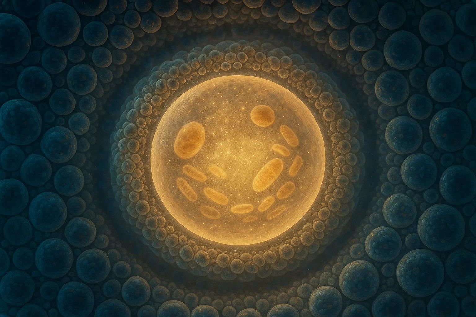
Human eggs, also known as oocytes, are amazing cells in that they are formed in the fetal development process and could lie dormant in the ovary for decades later as they only mature, then maybe fertilize. This longevity requires extraordinary protein and organelle cellular upkeep to secure quality and the fertility of the eggs.
The most recent landmark study by Zaffagnini et al. (2025) is to explore proteostasis (a balance between the levels of protein synthesis, folding and degradation) on a global scale in healthy human oocytes. The scientists tried it with more than 100 new donated eggs of healthy young women, and they discovered that these cells have surprising differences in the way they govern their protein and organelle activity versus those surrounding somatic cells and even mouse oocytes.
What is proteostasis and why is it significant?
Proteostasis is the term that denotes the cellular mechanisms that make sure the well-being and activity of the proteome, or every one of the proteins in a cell. The leaders are:
Proteasomes: Complexes of protein that break down worn-out or unnecessary proteins.
Lysosomes: Gallions that break down cellular wastes: heaps of proteins and ruined structures.
Mitochondria: Energy-producing cell organelles, which determine the metabolism of the cell.
Healthy cells require proper proteostasis. Protein aggregation, oxidative stress and cellular dysfunction may be the consequences of the disruptions, which are associated with aging and diseases.
Key Findings: The decrease in the Degradative Activity of the Human Oocytes
The study analyzed immature (germinal vesicle, GV) and mature (metaphase II, MII) oocytes of humans and their enclosed cumulus cells ( the follicle which helps the growth of the oocyte). The large scale findings were:
Oocytes possess a Low Level of Lysosomal and Proteasomal Activities compared to Somatic Cells
Experiments with fluorescent probes of lysosomal and proteasomal activity included that other researchers discovered that human oocytes have almost half the rates of the surrounding cumulus cells of degradation. This indicates that the process of protein degradation may not be much involved in the long period of, the dormant and maturation of oocytes.
Degradative Activity is further diminished upon Oocyte Maturity
In leading to a notable decrease in lysosomal and proteasomal activity in comparison with the immature GV-stage oocytes, there was a pronounced slowdown of the same in mature MII-stage oocytes. The above observation is contrary to what would be expected, that is increase in these activities in cellular renewal with maturation.
The increased Lysosomal Exocytosis in Mature Oocytes
Interestingly, as the amount of the intracellular lysosomes became low in mature oocytes, the study also noted an increase in the lysosomal proteins on the plasma membrane therefore implying that during maturation, the lysosomes are released outside the cell through lysosomal exocytosis.
The Mitochondrial Activity Goes Down Too with Maturation
Mitochondrial health and activity was indicated by the mitochondrial membrane potential that was lower in the mature oocytes than in the immature ones. Such a decline in the activity of mitochondria could be a way of avoiding oxidative damage that could arise as a result of long maturation.
There is Presence of Protein Agglomerates in Larger Lysosomes
As opposed to the case with mouse oocytes, whereby the protein aggregates are sequestered in special compartments known as ELVAs, human oocytes contain aggregates in a few large lysosomes known as refractile bodies. This variation illustrates the species-specific forms that are adjusted to proteostasis.
What Do These Findings Mean for Female Fertility?
The research hypothesis undermines the hypothesis that a slower organelle activity in human oocytes is part of adaptation because inhibited activity is essential to conserve major cellular components over the extended maturation process. Protein degradation and mitochondrial inhibition potentially limits some of the reactive oxygen species and other cellular stressors that form during development, and thus retains egg quality and the potential of eggs to develop.
This observation is crucial since:
It points out to an exceptional and human-specific proteostatic program that is not comparable to the model organisms such as mice.
It guides assisted reproductive technologies (ART) because it stresses that natural conditions surrounding oocytes should be imitated and this is of great importance because in vitro maturation frequently results in poor fertility performances.
It identifies possible oocyte quality biomarkers that concern lysosomal and mitochondrial activity.
Future Directions and clinical Implications
The authors suggest that future research should be done to:
Explain molecular processes that govern lysosomal exocytosis, and redistribution of organelles in oocytes.
Discuss the role of proteostatic adaptations in early embryonic processes of the embryo and its fertility.
Determine their relevance to aged or infertile oocytes and potential targets to enhance the success of ART.
Insight into the proteostasis peculiarities of the oocyte may become a step toward improved diagnostics and treatment of female infertility.
Conclusion: A Landmark Study in Reproductive Biology
The study by Zaffagnini et al. portrays a complete and novel picture of protein homeostasis in normal human oocytes. This study identifies a tightly balanced cellular approach that aids survival of the egg, through decades, by showing a reduction of the degradative and mitochondrial processes during the maturation.
Such perceptions further enlighten the molecular perception of female fertility and lead to a future that puts more fertility knowledge into practices that take the distinct biology of human oocytes into consideration.
Reference
Zaffagnini, G., Solé, M., Duran, J. M., Polyzos, N. P., & Böke, E. (2025). The proteostatic landscape of healthy human oocytes. The EMBO Journal.

