
The issue of targeting the drugs to reach the intended targets within the body is one of the greatest challenges in modern medicine. Although researchers have come up with effective medication in the treatment of cancer and other deadly diseases, tracing the exact location of the drugs has been a great challenge. The conventional imaging techniques are commonly dependent on fluorescent dyes or radioisotopes, yet these tracers may dissociate with the drug carriers and this results in misleading or inconclusive outcomes.
A group of researchers at Waseda University in Tokyo (collaborating with Osaka University and Kyoto University) have now announced a revolutionary new imaging method that addresses this issue. They have developed an approach, using activated gold nanoparticles (AuNPs), that enables real time tracing of the movement of drugs in the body without external tracers. This innovation may change the targeted drug delivery, particularly in cancer treatment, and become a gateway to highly specific effective treatment.
What is the Gold nanoparticles and Why are they important?
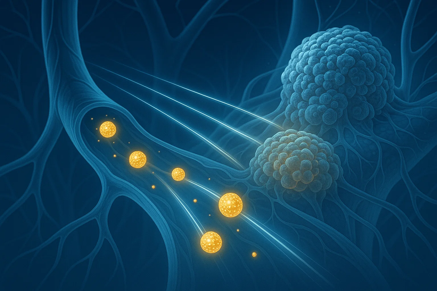
Gold nanoparticles are ultra-small particles of gold, most of the time between 1-100 nanometers. Although they have a very small size, they have distinctive chemical and biological characteristics. They hold promise especially in medicine since they are naturally concentrated in tumours. This qualifies them to be perfect drug delivery systems whereby the cancer drugs can be modified to attach to AuNPs and be transported to the tumor sites with reduced harm caused to the normal tissues.
The real challenge however has been monitoring their movement after they are in the body. Up to this point, researchers have used dyes or radioactive tracers to label the nanoparticles, yet these labels tended to detach with the AuNPs, resulting in low quality tracking drug distribution.
The Breakthrough: Neutron Activation Imaging
This problem is addressed using the new technique created by the Waseda team which directly changes the gold nanoparticles themselves. The scientists transformed stable gold atoms (197Au) to radioactive form (198Au) using a process known as neutron activation.
This change is important since 198Au produces gamma rays, which can be seen outside the body by utilizing specialized imaging devices. Notably, this modification does not affect the chemical properties of gold and thus the nanomaterials remain effective drug carriers.
Activation of atoms by irradiation with particles is a method that directly modifies the material. The modified elements release X-rays and gamma rays which cause the material to be visible outside the body without a degradation in its chemical properties.
Testing in Cancer Models
The researchers injected tumor carrying mice with radioactive AuNPs to test the technology. They could visualize the nanoparticles within the body over long durations giving it a clear and long term image of the movement and accumulation of the drug carriers within the tumors.
The paper has also discussed the application of this method to astatine-211 (211At) which is an effective radio therapeutic agent applicable in treatment of cancer. Although 211At is efficient, its half-life (7.2 hours) is short and causes it to be hard to detect after two days. When the researchers tagged the drug using 198Au nanoparticles, they were able to improve the tracking window to five days because 198Au has a longer half life of 2.7 days.
Why This Matters: Toward Better Cancer Treatment
This development may be a paradigm shift in specific drug delivery and cancer therapy. Allowing direct and long term monitoring of drugs within the body, doctors and researchers can understand as never before:
The manner of drug distribution within various tissues.
The duration of their effect at the site.
Whether they are successfully penetrating tumors.
How to maximize the effect of doses at a minimum side effect.
In addition, the strategy may result in safer drugs because real time pharmacokinetic studies, which is the study of drug movement in the body, can be done through this approach.
Discussing the Future: The Future of Nanomedicine
The research team is already intending on improving the technique further. They are concerned to enhance the imaging resolution and extend the methodology to other categories of nanoparticlebased drugs systems.
By further refining neutron activation imaging, we aim to make drug monitoring a clinical reality, potentially revolutionizing the field of imaging technologies.
When successful, this innovation will have the ability to revolutionize nanomedicine and lead to more accurate and personalized cancer and other treatment.
Conclusion
Real time repositioning of gold nanoparticles without the use of external tracers is a significant advance in drug delivery and medical imaging. This discovery by a group of researchers at Waseda University may make the future of cancer treatment different as drugs will be delivered to their target sites effectively and safely.
With gold nanoparticles pointing the path, the dream of smarter, more accurate and more personalized medicine is coming to life.
References & Resources
Koshikawa, N., Kataoka, J., Toyoshima, A., Kato, H., Kadonaga, Y. (2025). Activation imaging of gold nanoparticles for versatile drug visualization: An in vivo demonstration. Applied Physics Letters. DOI: 10.1063/5.0251048
Waseda University Press Release: Lighting the way: How activated gold nanoparticles reveal drug movement in the body
Phys.org coverage of the study: Gold Nanoparticles in Drug Delivery