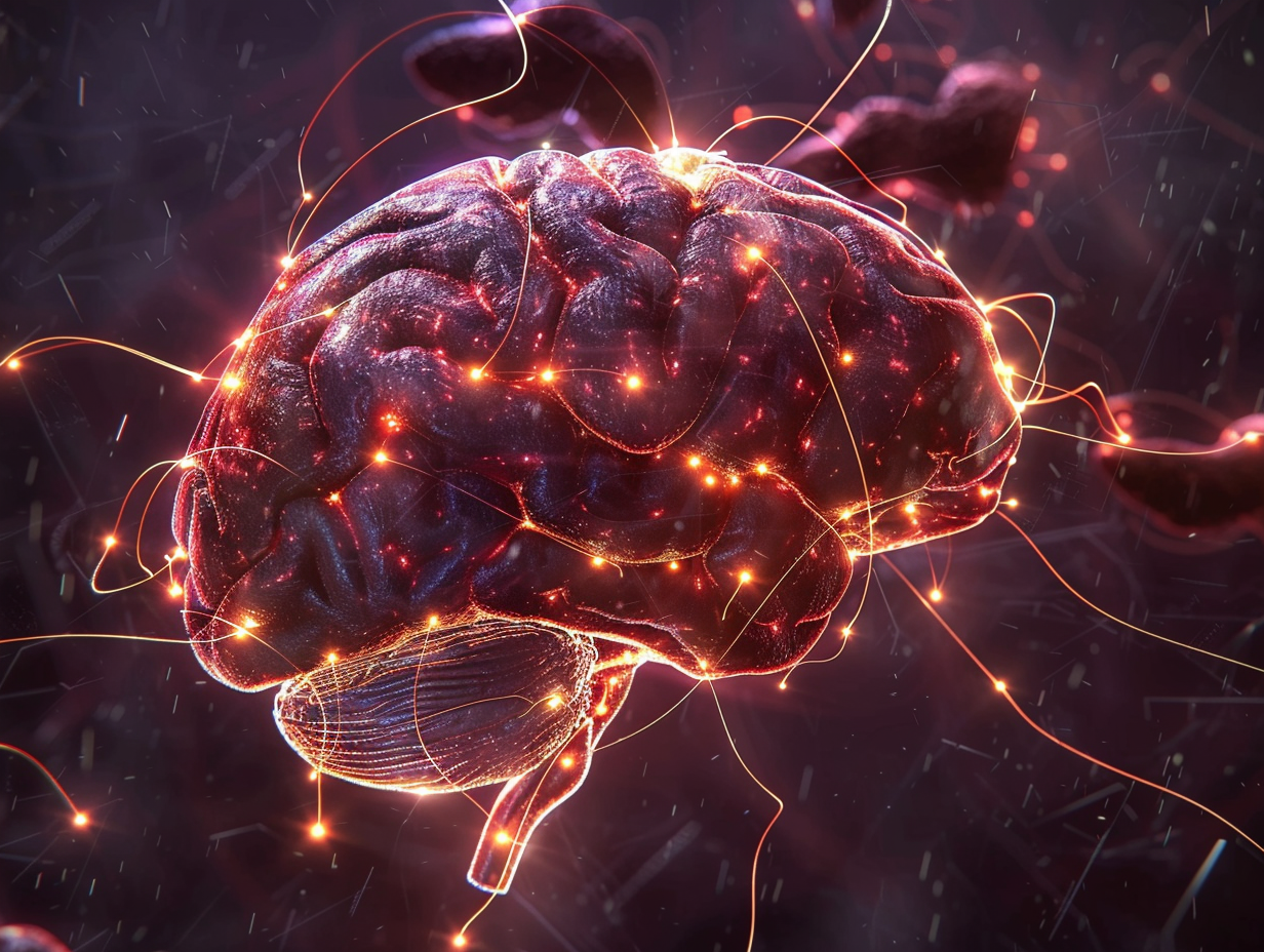
The majority believe that it is the role of pancreas, insulin and liver to control blood sugar levels. However, the brain is equally important in maintaining the level of glucose. Even decades ago, researchers were aware of the fact that brain circuits can be used to avoid excessively low blood sugar levels at the moment of danger, such as that of hypoglycemia. What has not been evident is the role of the brain in day to day glycemic control, particularly on a normal fasting basis, e.g. when we sleep at night without having eaten.
In a new study, published in Molecular Metabolism ( July 2025), it is found that a specific set of neurons in the ventromedial hypothalamus (VMH) are involved in regulating blood sugar in normal life. These are the cholecystokinin b receptor (Cckbr) marked neurons, which do not influence the appetite or body weight but have a distinctive role in glucose mobilization when fasting is short term.
Why This Matters
The balance of blood sugar or glucose homeostasis is crucial to survive. Inadequate glucose may negatively affect brain operation whereas excessive amounts will lead to diabetes and other complications.
The majority of the known hypothalamic neurons regulate the balance of energy (such as metabolism and appetite) and the level of glucose. This has led to confusion of the identification of neurons that can specifically control the glucose without having to change the food consumption or weight. The new VMH Cckbr neuron discovery provides a rare glimpse into a circuit in the brain that is devoted to glucose mobilization to the largest part.
The Study at a Glance
University of Michigan researchers and colleagues silenced or stimulated mice VMH Cckbr neurons and monitored the impacts on blood sugar during fasting and feeding cycles.
Silencing experiments: Mice experienced reduced blood sugar levels when such neurons were silenced during shorten fasts (e.g., the early light period when mice naturally eat less).
Activation experiments: The artificial stimulation of the neurons increased blood sugar rapidly with no alterations in liver glycogen breakdown or normal gluconeogenic gene activity.
Important process: These neurons activated lipolysis (breakage of fat) in white adipose tissue, which releases glycerol, a primary raw material to form new glucose in the liver, rather than drawing on the available stock of liver glycogen.

How VMH Cckbr Neurons Work
Mobilizing Fat for Sugar
Under normal conditions, glucose is produced in the liver in the state of fasting by breaking down glycogen or synthesis of new glucose using substances such as lactate, amino acids, and glycerol. The researchers discovered that VMH Cckbr neurons enhance the generation of glucose through the increased release of glycerol on fat tissue.
This occurs via the sympathetic nervous system (SNS) and is reliant upon b3- adrenergic receptors in fat cells. The blockage of these receptors eliminated the glycerol release (and subsequent glucose increase) .
The Not Your Average Glucose Circuit.
Interestingly, such neurons fail to activate the typical postulates experienced in acute hypoglycemia. They:
Do not take more glucagon (an insulin-like hormone).
Do not cause liver glycogen depletion.
Do not boot up gluconeogenic genes, such as PEPCK or G6Pase.
Instead they are a specialized circuit that optimizes glucose levels during normal fasting, providing adaptability in accordance with the body requirements.
The importance of this discovery.
For Diabetes Research
Overproduction of glucose and harmful accumulation of ketones are caused by excessive fat breakdown in type 1 diabetes. Learning about the mechanisms of brain pathways such as VMH Cckbr neurons to regulate lipolysis may result in interventions to eliminate dangerous fluctuations in sugar levels in the blood.
To prevent Hypoglycemia.
Hypoglycemia is a significant risk in individuals taking insulin who have diabetes. Improving or safeguarding these neurons could be of benefit to patients to evade low sugar outbursts through enhanced natural glucose mobilization.
In the case of Obesity and Metabolic Disease.
Too much SNS induced lipolysis has been associated with insulin resistance and fatty liver disease. When VMH Cckbr neurons are hyperactive in the state of obesity, then they may be contributing to metabolic dysfunction.
Looking Ahead
The research also poses some new intriguing questions:
What are the normal signals that cause the activation of these neurons in the normal fasting condition?
Are their effects similar to those of drugs that act on the b3 adrenergic pathway?
Are human beings the same in their neuronal system and how it may vary in diabetes or obesity?
Although the study was performed on mice, the comparison with the physiology of humans is consistent enough to provoke the desire to conduct future clinical trials.
Conclusion
This is an innovative research that points at the fact that the brain actively helps to maintain the level of blood sugar in everyday life not only in crisis. VMH Cckbr neurons mobilize glycerol stored in fat tissue providing the liver with the fuel it requires to maintain glucose levels constant during brief periods of starvation.
The finding highlights a fundamental teaching, which is as follows: the brain is not a passive observer of metabolism, but an active controller of blood sugar. With more information about these circuits being revealed, there are new therapeutic opportunities now in diabetes, hypoglycemia, and other metabolic diseases.
Reference
Su J, Hashsham A, Kodur N, et al. Control of physiologic glucose homeostasis via hypothalamic modulation of gluconeogenic substrate availability. Molecular Metabolism. 2025;99:102216. doi:10.1016/j.molmet.2025.102216

