
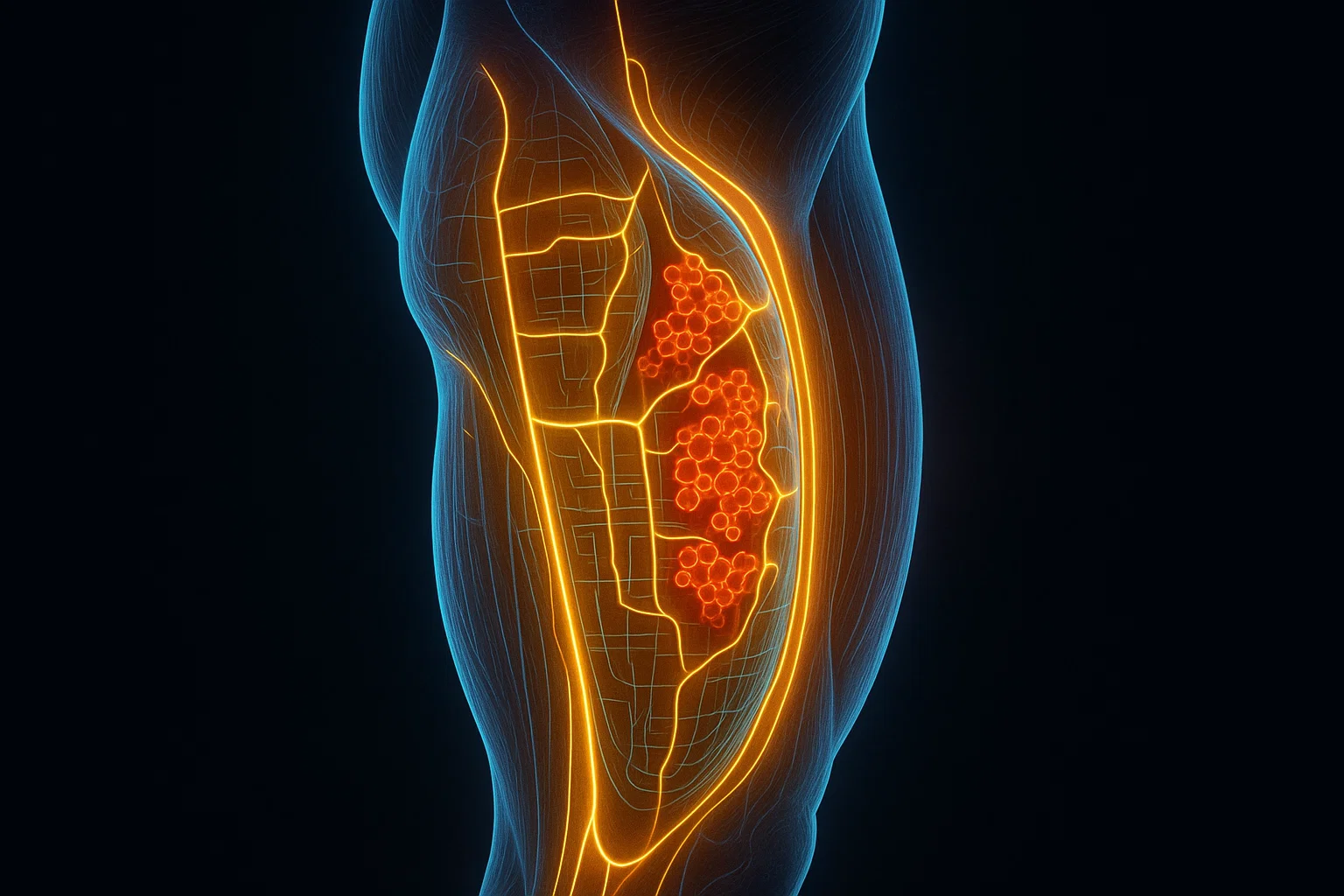

What if understanding type 2 diabetes required looking beyond blood sugar and examining the molecular activity inside our muscles? Scientists have developed detailed maps of skeletal muscle proteins that reveal previously hidden patterns of insulin resistance. New 2026 findings offer further clues about how muscle metabolism contributes to diabetes and how these discoveries might eventually support more personalized prevention and treatment.
Skeletal muscle plays a central role in regulating blood sugar. It is responsible for much of the body’s insulin-stimulated glucose uptake.
A 2025 study in Cell analyzed skeletal muscle samples from 123 participants and found molecular patterns strongly associated with insulin sensitivity.
The researchers identified protein and protein-phosphorylation signatures that helped estimate whole-body insulin sensitivity, even when ordinary blood sugar measurements did not capture the full differences.
Research published in 2026 uncovered additional muscle-cell mechanisms, including the potential role of mitochondrial protein Miro1 in insulin resistance.
These molecular maps are research tools, not routine clinical tests. They have not yet been shown to predict future diabetes or determine the best treatment for individual patients.
Established preventive approaches still matter. Regular physical activity, healthy eating, appropriate weight management, and evidence-based medical care can substantially reduce the risk of type 2 diabetes.
Why Scientists Are Looking Beyond Blood Sugar
For decades, type 2 diabetes diagnosis and management have relied heavily on blood glucose measurements.
Tests such as fasting plasma glucose, the oral glucose tolerance test, and hemoglobin A1c (HbA1c) help doctors determine whether someone has normal glucose levels, prediabetes, or diabetes.
These measurements are essential, but they do not explain everything happening inside the body.
Two people can have similar blood glucose results while experiencing very different biological changes.
One person may have substantial insulin resistance but maintain relatively normal glucose levels because the pancreas produces additional insulin.
Another may have elevated glucose levels partly because the pancreas cannot produce enough insulin to compensate.
This difference matters because type 2 diabetes is not a single, uniform metabolic condition.
It develops through interacting changes involving skeletal muscle, the liver, fat tissue, pancreatic beta cells, and other organs.
Researchers are therefore investigating whether molecular measurements can reveal insulin resistance earlier and more precisely than conventional glucose measurements alone.
A major 2025 study published in Cell suggested that skeletal muscle contains valuable information about these metabolic differences.
The findings are part of a growing effort to develop precision medicine for type 2 diabetes, in which prevention and treatment strategies could eventually reflect an individual’s specific biological characteristics.
Why Skeletal Muscle Matters in Type 2 Diabetes
Skeletal muscle is best known for movement, strength, and physical activity.
However, it is also one of the body’s most important metabolic tissues.
After a meal, blood glucose rises, and the pancreas releases insulin.
Insulin signals muscle cells to take up glucose from the bloodstream and use or store it.
A transporter protein called GLUT4 plays a central role in this process.
When insulin signaling works normally, GLUT4 moves toward the muscle-cell membrane, allowing glucose to enter the Cell.
Once inside, cells can use glucose to generate energy or store it as glycogen.
What Happens During Insulin Resistance?
In insulin-resistant muscle, cells become less responsive to the effects of insulin.
As a result, glucose uptake may become impaired.
The pancreas often compensates by producing more insulin.
Over time, if pancreatic insulin secretion cannot adequately compensate for insulin resistance, blood glucose levels can rise and type 2 diabetes may develop.
Insulin resistance can appear years before diabetes is diagnosed, although not everyone with insulin resistance progresses to the disease.
Because skeletal muscle accounts for a large share of insulin-stimulated glucose disposal, changes in its molecular activity matter.
This makes muscle tissue a valuable target for understanding why insulin resistance develops and why people respond differently to lifestyle changes and medication.
The 2025 Cell Study: Mapping the Molecular Signatures of Insulin Resistance
The research that inspired this article was published online on May 27, 2025, and appeared in the July 24, 2025 issue of Cell.
Its title was:
“Personalized molecular signatures of insulin resistance and type 2 diabetes.”
The study was led by Jeppe Kjærgaard and colleagues, including scientists affiliated with the University of Copenhagen, Karolinska Institutet, and collaborating research institutions.
The researchers investigated whether skeletal muscle’s molecular composition could reveal differences in insulin sensitivity between people with normal glucose tolerance and those with type 2 diabetes.
Rather than analyzing only blood sugar levels, the team examined thousands of proteins and their chemical modifications within muscle tissue.
Original research: Kjærgaard et al., Cell (2025).
PubMed record: PMID 40436015.
How Was the Study Conducted?
The study combined a discovery cohort of 77 participants with an additional validation cohort of 46 participants.
Together, the researchers examined data from 123 men and women.
The participants included people with normal glucose tolerance and people diagnosed with type 2 diabetes.
Researchers obtained samples from the vastus lateralis, a skeletal muscle in the thigh.
Researchers collected muscle biopsies under fasting conditions and during a controlled insulin infusion.
The investigators used advanced laboratory techniques to analyze two important molecular layers.
Proteomics: The study of proteins present in cells or tissues, including differences in protein abundance.
Phosphoproteomics: The study of proteins modified through phosphorylation, a chemical process that helps regulate many cellular signaling pathways.
By combining these measurements with detailed metabolic testing, the scientists created a molecular map of how skeletal muscle differs across varying degrees of insulin sensitivity.
What Did the Muscle Protein Maps Reveal?
The study identified several important findings that deepen our understanding of type 2 diabetes.
1. Muscle Protein Patterns Were Strongly Associated With Insulin Sensitivity
One central finding was that the molecular composition of skeletal muscle could help estimate a person’s insulin sensitivity.
The researchers compared muscle protein measurements with insulin sensitivity assessed using a technique called the hyperinsulinemic-euglycemic clamp.
This method involves administering insulin while carefully adjusting a glucose infusion to maintain blood sugar within a target range.
The amount of glucose needed to maintain that level helps researchers assess the body’s response to insulin.
Although the procedure is highly informative, it is labor-intensive and not routinely used for diabetes screening.
The scientists found that patterns in both the muscle proteome and phosphoproteome were strongly associated with clamp-measured insulin sensitivity.
These associations provided information that was not fully captured by fasting glucose or HbA1c measurements.
Why this matters: A person’s blood sugar result may not fully reflect the molecular changes already occurring in tissues involved in insulin action.
However, identifying an association with current insulin sensitivity is different from predicting future diabetes risk.
The research did not establish a clinically validated test to predict who will develop diabetes.
2. Fasting Muscle Samples Contained Valuable Information
Another interesting finding was that muscle samples obtained before insulin stimulation contained molecular signatures associated with whole-body insulin sensitivity.
This suggests that insulin sensitivity differences are reflected not only in the muscle’s immediate response to insulin, but also in its underlying molecular state.
The study showed that fasting muscle proteome and phosphoproteome patterns were informative about insulin sensitivity.
This could eventually help researchers identify biological pathways associated with insulin resistance without relying exclusively on measurements collected during acute insulin stimulation.
Nevertheless, routine clinical application would require further validation and a practical, accurate testing approach.
3. Not Every Insulin Signaling Pathway Was Impaired
Insulin resistance is sometimes described as though the entire insulin signaling system stops functioning.
The 2025 research revealed a more complicated picture.
The researchers identified molecular signaling processes that were disrupted in insulin-resistant muscle, alongside others that remained relatively preserved.
This phenomenon is sometimes called selective insulin resistance.
In other words, different cellular responses to insulin may be affected to different degrees.
Understanding these distinctions could inform future therapies.
Instead of assuming all insulin-related signaling is equally impaired, researchers may eventually identify specific molecular processes to target more precisely.
However, the study did not test a new drug or show that treating one of these pathways improves patient outcomes.
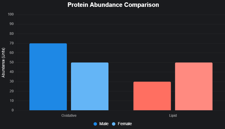
4. Men and Women Showed Differences—and Important Similarities
The researchers also investigated differences between male and female participants.
They observed substantial sex-related variation in skeletal muscle protein abundance and phosphorylation patterns.
These differences align with the broader understanding that muscle metabolism varies between individuals.
Surprisingly, however, many of the molecular signatures associated with insulin resistance were similar in men and women.
This suggests that although baseline muscle biology can differ, some insulin-resistance mechanisms may be shared across sexes.
Future research will need to determine whether these observations affect treatment responses or support different prevention strategies.
What Is a Muscle Molecular Map?
The term muscle map does not refer to a conventional medical image.
It describes a detailed collection of molecular measurements that researchers can use to understand cellular biology.
For example, a molecular muscle map may include measurements of protein abundance, protein modifications, metabolic pathways, and patterns of cellular signaling.
Researchers can analyze these measurements together to identify biological features associated with insulin resistance.
| Measurement | What researchers learn |
|---|---|
| Blood glucose | How much glucose is circulating in the bloodstream |
| HbA1c | Average blood glucose exposure over roughly two to three months |
| Insulin sensitivity clamp | How effectively the body responds to insulin under controlled conditions |
| Muscle proteomics | Which proteins are present and how their abundance varies |
| Muscle phosphoproteomics | How protein signaling activity differs across metabolic states |
| Single-nucleus transcriptomics | Which genes are active in different muscle-cell nuclei and associated cell populations |
These approaches offer complementary information.
None should be considered a complete replacement for established clinical testing.
How Could Muscle Mapping Support Personalized Diabetes Prevention?
The ultimate goal of precision medicine is to understand why individuals develop disease differently and use that knowledge to improve care.
In type 2 diabetes, this could involve identifying particular molecular pathways that contribute to insulin resistance in different people.
Consider two hypothetical individuals with similarly elevated HbA1c levels.
One might have pronounced abnormalities in muscle glucose transport, while another might have different changes involving mitochondrial metabolism, inflammation, or pancreatic insulin secretion.
A well-validated molecular testing approach could help researchers distinguish these biological patterns.
In the future, this information might assist with:
- Identifying metabolic abnormalities that conventional glucose measurements do not fully capture.
- Developing treatments directed toward specific mechanisms of insulin resistance.
- Understanding why individuals respond differently to exercise, diet, or medication.
- Investigating biological changes before diabetes becomes clinically apparent.
- Designing more targeted clinical trials.
However, these applications are still largely research goals.
The 2025 Cell study did not demonstrate that muscle profiling can prevent diabetes or select an effective personalized treatment for a patient.
Before such testing could enter routine care, scientists would need prospective studies, independent validation, clinically useful thresholds, and evidence that testing improves health outcomes.
What New Research in 2026 Has Revealed
Since the original muscle-mapping study, several investigations have expanded our understanding of the relationship between skeletal muscle and insulin resistance.
These studies do not validate the 2025 protein map as a clinical diagnostic tool, but they strengthen the case for investigating muscle biology at a more detailed molecular level.
February 2026: A New Single-Nucleus Atlas of Muscle Insulin Resistance
A February 2026 preprint investigated insulin resistance at the level of individual nuclei in human skeletal muscle.
The researchers analyzed 25,303 nuclei obtained from muscle samples involving 18 older adults with obesity, including participants with and without type 2 diabetes.
Using single-nucleus RNA sequencing, they identified distinct molecular populations within skeletal muscle.
Some populations showed gene-expression patterns associated with more favorable insulin sensitivity, while others were associated with impaired metabolic function.
The researchers also investigated candidate mechanisms involving the zinc transporter ZIP14 and epidermal growth factor signaling.
The findings suggest that different populations of muscle-cell nuclei may contribute differently to insulin resistance.
This is important because skeletal muscle is not a uniform collection of identical cells.
Different cell types and different nuclei within muscle fibers can have specialized biological roles.
Research source: A full-length single-nucleus transcriptomic atlas of human skeletal muscle insulin resistance (2026).
Important limitation: This work was available as a preprint, meaning it should not be treated as equivalent to a fully peer-reviewed clinical validation study. The initial participant sample was also relatively small.
September 2026: Muscle Cells May Influence Other Organs
Research highlighted by Joslin Diabetes Center in September 2026 investigated molecular differences between muscle cells derived from people with and without type 2 diabetes.
Scientists used induced pluripotent stem cells to grow muscle cells in laboratory conditions, allowing them to study characteristics that might persist outside the original diabetic environment.
The researchers examined gene expression, proteins, and molecules the cells release.
This approach may help clarify whether some differences in muscle metabolism are intrinsic to the cells rather than simply consequences of elevated blood glucose.
The findings suggest that muscle tissue may contribute to metabolic disease not only through glucose uptake, but also through molecular communication with other organs.
Institutional research summary: Joslin Diabetes Center, September 14, 2026.
October 2026: Scientists Identify a Role for Miro1
One of the most recent developments came from a study published in Nature Communications on October 19, 2026.
The study, titled “Miro1 mediates skeletal muscle insulin resistance in type 2 diabetes,” investigated a protein involved in mitochondrial regulation.
Mitochondria are cellular structures that help generate energy.
Their organization and function are important for muscle metabolism.
The researchers found that a protein called mitochondrial Rho GTPase 1, or Miro1, accumulated in skeletal muscle from people with obesity and type 2 diabetes.
Additional experiments suggested that abnormal Miro1 regulation could contribute to impaired insulin action.
Importantly, the investigation also included data from exercise training.
Exercise training was associated with reduced skeletal muscle Miro1 accumulation and improved insulin sensitivity in people with type 2 diabetes.
In mice and laboratory cell models, removing Miro1 from muscle cells improved several measures related to mitochondrial function and glucose metabolism.
These findings identify Miro1 as a potential research target for understanding and treating insulin resistance.
However, the study does not establish that Miro1-targeting medicines are safe or effective in humans.
Original research: Heintz et al., Nature Communications, October 19, 2026.
Why Exercise Remains So Important
New research on Miro1 offers another possible explanation for why exercise can improve insulin sensitivity.
Physical activity influences multiple biological processes in skeletal muscle.
Regular exercise can improve glucose transport, mitochondrial function, insulin signaling, and other aspects of metabolic health.
Some benefits occur even without substantial weight loss.
The October 2026 Miro1 study strengthens the molecular evidence linking exercise training to improvements in muscle metabolism.
However, the practical recommendation to remain physically active does not depend on this newly identified protein.
Extensive clinical evidence already supports it.
The American Diabetes Association’s 2026 Standards of Care recommend structured lifestyle interventions for adults at high risk of type 2 diabetes.
For adults with overweight or obesity at elevated risk, these programs typically target at least 150 minutes of moderate-intensity physical activity per week and, when appropriate, a sustained weight loss of about 5–7%.
The U.S. Diabetes Prevention Program demonstrated that intensive lifestyle intervention reduced the incidence of type 2 diabetes by approximately 58% among high-risk participants during the original trial period.
These results show that meaningful prevention is possible without requiring experimental muscle biopsies or molecular profiling.
Clinical guidance: American Diabetes Association Standards of Care in Diabetes—2026.
Public health resource: CDC National Diabetes Prevention Program.
Can Muscle Mapping Predict Diabetes Before Blood Sugar Rises?
This is one of the most exciting possibilities, but the evidence needs careful interpretation.
The 2025 study showed that muscle protein patterns were associated with insulin sensitivity among the people examined.
That is not the same as demonstrating that those patterns can predict future diabetes.
To establish predictive usefulness, researchers would need to measure muscle signatures in people without diabetes, follow them over time, and determine which individuals later develop the disease.
A clinically useful test would also need to demonstrate meaningful accuracy beyond existing risk factors and screening tools.
Researchers would need to evaluate whether its benefits justify the cost and invasiveness of muscle biopsy.
For now, established methods such as HbA1c, fasting glucose, and oral glucose tolerance testing remain central to diabetes diagnosis and risk assessment.
Molecular muscle maps are not ready to replace them.
What Are the Main Limitations of the Research?
Although the 2025 Cell study and subsequent investigations provide valuable information, several limitations deserve attention.
Relatively Small Research Cohorts
The original study included 123 participants across two cohorts.
This is a meaningful sample for an intensive muscle proteomics investigation, but much larger studies would be needed to establish population-wide clinical reliability.
Association Does Not Establish Causation
A protein pattern associated with insulin resistance may contribute to the condition, result from it, or reflect another biological process.
Further experiments are needed to determine which molecular changes directly influence insulin sensitivity.
Muscle Biopsies Are Invasive
Collecting muscle tissue requires a medical procedure.
Even if the molecular findings prove clinically valuable, widespread testing would require practical, safe, and cost-effective implementation.
Diabetes Is a Multi-Organ Disease
Skeletal muscle is extremely important, but type 2 diabetes also involves the pancreas, liver, adipose tissue, and other organs.
A molecular muscle map cannot fully explain all the biological mechanisms underlying the disease.
Personalized Treatments Have Not Been Validated
The researchers did not conduct a clinical trial in which treatments were selected according to individual muscle protein signatures.
Therefore, there is no established evidence that this method improves outcomes compared with standard care.
Diverse Populations Need Further Study
Researchers should validate the findings across different ages, backgrounds, levels of physical activity, and metabolic conditions.
The results of specialized research cohorts may not apply equally to every patient.
What Can People Do Today to Reduce Type 2 Diabetes Risk?
While scientists investigate the future of precision medicine, people at risk of type 2 diabetes can already benefit from evidence-based preventive strategies.
Stay Physically Active
Regular walking, cycling, swimming, and other aerobic activities can support insulin sensitivity and cardiovascular health.
Resistance training can also support muscle strength and glucose regulation.
Adapt activity plans to individual health status and physical ability.
Follow a Sustainable Healthy Eating Pattern
A balanced eating pattern that emphasizes vegetables, appropriate portions of fruit, whole grains, legumes, and other nutrient-rich foods can support metabolic health.
Reducing excessive intake of sugary drinks and highly processed foods may also help.
No single diet is appropriate for everyone.
Manage Weight Where Appropriate
For some people with overweight or obesity and elevated diabetes risk, modest sustained weight loss can improve insulin sensitivity and reduce the likelihood of developing type 2 diabetes.
Individualize weight management, especially for people with existing medical conditions.
Get Recommended Screening
People with diabetes risk factors should discuss appropriate screening with a healthcare professional.
Risk factors may include a family history of diabetes, excess abdominal fat, previous gestational diabetes, and certain metabolic conditions.
Follow Individualized Medical Advice
People who already have prediabetes or diabetes may benefit from structured lifestyle programs, medication, and monitoring according to their circumstances.
New molecular discoveries should complement, not replace, established care.
Conclusion: A New Understanding of Diabetes Is Emerging From Our Muscles
The 2025 Cell study marked an important advance in understanding the molecular biology of insulin resistance.
By combining detailed skeletal muscle proteomics with direct measurements of insulin sensitivity, researchers identified patterns that conventional blood sugar tests do not fully reveal.
The findings demonstrated that insulin resistance involves diverse molecular changes and that skeletal muscle contains valuable clues about metabolic health.
New studies published in 2026 have expanded this picture.
Single-nucleus mapping has revealed additional complexity within muscle tissue, while research into Miro1 and mitochondrial regulation has identified potentially important mechanisms linking exercise and insulin sensitivity.
Together, these discoveries could eventually support more targeted approaches to preventing and treating type 2 diabetes.
However, they have not yet produced a validated muscle-based diagnostic test or a proven personalized prevention program.
The real breakthrough is not that scientists can now predict diabetes from a muscle map, but that they are beginning to understand insulin resistance in far greater molecular detail.
That understanding may lead to better treatments and earlier interventions in the future.
Until then, established screening, regular physical activity, healthy eating, and appropriate medical care remain the most reliable tools for reducing diabetes risk.
References
1. Kjærgaard, J., et al. (2025). Personalized molecular signatures of insulin resistance and type 2 diabetes. Cell, 188(15), 4106–4122.e16.
https://doi.org/10.1016/j.cell.2025.05.005
2. Kjærgaard et al. (2025). PubMed record of the original muscle proteomics study.
https://pubmed.ncbi.nlm.nih.gov/40436015/
3. Whytock, K. L., et al. (2026). A full-length single-nucleus transcriptomic atlas of human skeletal muscle insulin resistance. bioRxiv preprint.
https://doi.org/10.64898/2026.02.26.708250
4. Heintz, E. C., et al. (2026). Miro1 mediates skeletal muscle insulin resistance in type 2 diabetes. Nature Communications, 17, Article 10702.
https://doi.org/10.1038/s41467-026-77278-6
5. Joslin Diabetes Center (2026). New Research Reveals Surprising Role for Skeletal Muscle in Type 2 Diabetes.
https://joslin.org/news-stories/all-news-stories/news/2026/09/new-research-reveals-surprising-role-for-skeletal-muscle-type-2-diabetes
6. American Diabetes Association (2026). Prevention or Delay of Diabetes and Associated Comorbidities: Standards of Care in Diabetes—2026. Diabetes Care.
https://doi.org/10.2337/dc26-s003
7. Centers for Disease Control and Prevention. Preventing Type 2 Diabetes With the Lifestyle Change Program.
https://www.cdc.gov/diabetes-prevention/lifestyle-change-program/