
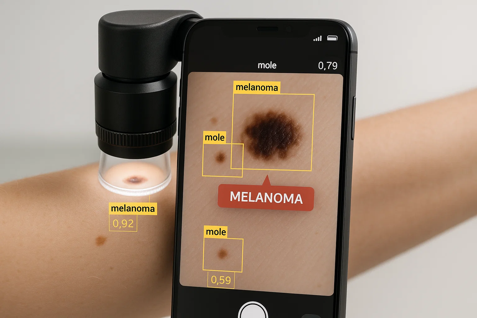

Skin cancer is among the most common forms of cancer worldwide. Detecting suspicious skin changes early can make treatment more effective, particularly for melanoma, a potentially life-threatening cancer that can spread to other organs.
However, identifying a dangerous lesion is not always straightforward.
Some melanomas resemble ordinary moles, while harmless skin growths can display features that raise concern. Dermatologists use clinical examinations, dermoscopy, medical history, and sometimes biopsies to distinguish between these conditions.
Artificial intelligence (AI) is emerging as a potentially valuable tool in this process.
Through analyzing thousands or millions of skin images, deep-learning systems can learn visual patterns associated with different types of skin lesions. Researchers are investigating whether these systems can help healthcare professionals recognize suspicious changes, prioritize examinations, and boost diagnostic consistency.
Two important studies published in 2024 and 2025 exhibited promising results using convolutional neural networks (CNNs) for skin lesion classification.
More recent research involving advanced dermatology core models has expanded these possibilities, showing that AI can analyze multiple image types and potentially improve clinician performance.
Yet an essential distinction remains: strong accuracy in an experimental dataset does not automatically mean an AI system is safe or reliable enough to diagnose skin cancer in real patients.
This article examines the science behind AI skin lesion analysis, the evidence supporting its potential, and the challenges that must be addressed before widespread clinical integration.
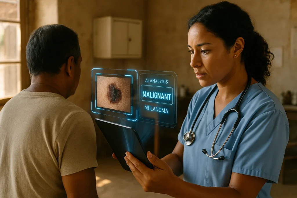
Why Early Skin Cancer Detection Matters
Skin cancer develops when abnormal skin cells proliferate uncontrollably.
Ultraviolet radiation from sunlight and tanning devices is a major preventable risk factor for several types of skin cancer.
The three principal types are:
Melanoma
Melanoma develops from melanocytes, the cells that produce the pigment melanin.
Even though less common than basal cell and squamous cell carcinomas, melanoma accounts for a disproportionate share of skin cancer deaths because it can spread to other parts of the body.
Early detection and appropriate treatment substantially improve the outlook for many patients.
Basal Cell Carcinoma
Basal cell carcinoma is a common type of skin cancer that typically develops in sun-exposed areas.
It usually grows slowly and rarely spreads to distant organs, but it can damage nearby tissues if left untreated.
Squamous Cell Carcinoma
Cutaneous squamous cell carcinoma develops from squamous cells in the skin.
It is often associated with cumulative ultraviolet exposure and can occasionally spread beyond its original location.
All three conditions require appropriate medical assessment and treatment.
Skin Cancer Statistics in 2026
According to the US National Cancer Institute’s Surveillance, Epidemiology, and End Results (SEER) program, an estimated 112,000 new invasive melanoma cases and 8,510 melanoma deaths are expected in the United States during 2026.
The five-year relative survival rate for localized melanoma is reported at approximately 100%, compared with approximately 34% for melanoma diagnosed after distant spread.
These are population-level estimates, not predictions for an individual patient.
They show why identifying and treating melanoma early matters.
Source: National Cancer Institute, Melanoma of the Skin: Cancer Stat Facts
The Challenge of Diagnosing Skin Lesions
Dermatologists examine skin lesions using visual patterns, medical history, and, when appropriate, specialized imaging.
One commonly used technique is dermoscopy.
A dermatoscope provides magnified views of skin structures that may be difficult to evaluate with the naked eye.
Dermatologists examine features such as:
- Asymmetry and irregular lesion shape.
- Uneven borders.
- Variation in pigmentation.
- Changes in size or appearance.
- Unusual internal structures.
- Patterns of blood vessels.
- Signs of bleeding, ulceration, or other concerning changes.
Despite advances in dermatology, several difficulties remain.
Visual similarities: Benign and malignant lesions sometimes look similar.
Diagnostic variability: Clinicians may interpret some borderline or unusual lesions differently.
Large image collections: Monitoring patients with many moles can require reviewing numerous images over time.
Access to specialists: Dermatology services may be limited in rural or underserved communities.
Rare presentations: Less common skin cancers or atypical appearances can be more difficult to recognize.
Artificial intelligence could potentially help address some of these problems, particularly by locating patterns in large image datasets and assisting clinicians during assessment.
However, an AI prediction cannot replace the complete clinical evaluation.
The 2025 Research Breakthrough: An Optimized CNN for Melanoma Detection
A study published on July 1, 2025, in Scientific Reports examined a deep-learning approach to detect melanoma in dermoscopic images automatically.
The research, conducted by Vida Esmaeili, Mahmood Mohassel Feghhi, and Hadi Seyedarabi, was titled:
Automatic melanoma detection using an optimized five-stream convolutional neural network.
The researchers endeavored to address several problems that commonly affect automated melanoma detection, including:
- Imbalanced image datasets.
- Differences in lesion size, shape, and orientation.
- Visual similarities between malignant and benign lesions.
- Hair and other image artifacts.
- Variations in color, texture, and image quality.
To confront these challenges, they combined image preprocessing, specialized feature extraction, and an optimized convolutional neural network.
What Makes the System Different?
Instead of relying only on general visual patterns, the approach processes several kinds of lesion information.
These include color, edges, texture, spatial characteristics, and directional features.
The researchers also used techniques designed to make the system less sensitive to image orientation and other variations.
The study reported excellent performance across several public skin lesion image datasets.
However, the findings were based on computational evaluations rather than a prospective trial showing improved clinical results in practice.
Original study: Esmaeili et al., Scientific Reports (2025)
How AI Skin Lesion Analysis Works
AI-based skin lesion classification generally includes several connected processes.
Step 1: Collect Skin Images
The first step is obtaining images of suspicious skin lesions.
Researchers may use clinical photographs or dermoscopic images.
Clinical photographs show a lesion and its surrounding skin, while dermoscopic images uncover additional skin structures through magnification and controlled illumination.
Public research databases such as HAM10000 and the International Skin Imaging Collaboration (ISIC) archive provide important resources for developing computer-vision systems.
However, photographs taken under regulated conditions may differ substantially from images captured with ordinary smartphones.
Research resource: International Skin Imaging Collaboration
Step 2: Prepare the Images
Raw photographs may contain factors that interfere with automated interpretation.
These include hair, shadows, uneven lighting, image noise, and changes in camera angle.
Image preprocessing can involve resizing, color normalization, contrast enhancement, and artifact reduction.
The 2025 study used techniques including hair removal, image enhancement, and CNN-based denoising.
Such processing can help models focus on lesion characteristics rather than irrelevant visual interference.
Step 3: Address Dataset Imbalance
In medical imaging datasets, benign lesions often greatly outnumber malignant lesions.
This creates a challenge because a model could achieve deceptively high overall accuracy by favoring the most common category.
In 2025, researchers explored a generative adversarial network (GAN)-based approach to balance the images used in their research.
A GAN is a machine-learning system that can generate synthetic examples resembling its training data.
Synthetic images may help expand training data sets, but they must be evaluated carefully.
Poorly generated or unrealistic examples can cause artifacts and potentially affect model dependability.
Step 4: Extract Important Visual Features
Convolutional neural networks learn patterns from images through layers of mathematical operations.
In skin lesion analysis, these patterns may include:
- Lesion borders.
- Pigment distribution.
- Texture irregularities.
- Local changes in shape.
- Spatial relationships between image features.
The 2025 system used multiple specialized feature-extraction techniques to characterize lesion patterns.
Rather than interpreting every feature in isolation, the model combines information to distinguish between image categories.
Step 5: Classify the Lesion
After processing the image, the AI generates a classification or set of prediction scores.
For example, a model designed to distinguish melanoma from benign lesions may assign a score indicating how strongly an image resembles melanoma examples in its training data.
An important limitation is that a model’s prediction score is not necessarily the real-world probability that a patient has cancer.
Reliable probability estimates require appropriate calibration and testing in representative clinical populations.
Step 6: Support Clinical Assessment
In a properly validated clinical workflow, an AI result may provide additional information to healthcare professionals.
A dermatologist can consider that information alongside the patient’s history, examination findings, and other medical evidence.
When cancer is suspected, a biopsy with microscopic examination may be needed to establish the diagnosis.
What Accuracy Did the 2025 Study Report?
The researchers evaluated their proposed method using multiple established image datasets.
The published paper reported the following melanoma detection performance.
| Research dataset | Reported detection result |
| HAM10000 | 99.8% |
| ISIC 2024 | 99.9% |
| ISIC 2017 | 99.62% |
| ISIC 2016 | 99.6% |
These results were unusually high and suggest that the system performed strongly under the evaluation conditions used by the researchers.
However, several factors warrant careful interpretation.
First, dataset-level performance may not reflect what happens when a model encounters images from a different hospital, camera, or population.
Second, the balance of melanoma and benign lesions in research datasets can differ from their distribution among patients attending routine clinics.
Third, accuracy alone is insufficient to establish whether the technology is medically safe.
Researchers also need to investigate sensitivity, specificity, false-negative rates, calibration, and clinical consequences.
Finally, the reported results do not establish that the model would detect 99.8% or more of all melanomas in everyday healthcare settings.
The findings support further investigation rather than immediate reliance on the system for independent diagnosis.
The Earlier 2024 Study: Classifying Multiple Skin Lesion Types
A second study, published in Cluster Computing on June 17, 2024, investigated a deep convolutional neural network for multiclass skin cancer classification.
Essam H. Houssein and colleagues conducted the research.
Unlike a system focused primarily on melanoma detection, this study examined the ability of a network to distinguish between multiple skin lesion categories.
The researchers evaluated their model using the HAM10000 and ISIC 2019 datasets.
They also compared performance against multiple established deep-learning architectures, including VGG16, VGG19, DenseNet121, DenseNet201, and MobileNetV2.
Reported Performance
| Dataset | Reported classification accuracy |
| HAM10000 | 98.5% |
| ISIC 2019 | 97.1% |
The study reported that the proposed network outperformed the comparison models in its experimental assessments.
However, these results should not be directly compared with the percentages from the 2025 melanoma-focused study.
The two investigations used different architectures, datasets, evaluation procedures, and classification objectives.
A model distinguishing several lesion categories faces a different task from one focused on melanoma detection.
The 2024 article also received a published correction in March 2026, which readers should consult when evaluating the original research.
Original paper: Houssein et al., Cluster Computing (2024)
A Major Development in 2025: PanDerm and Multimodal AI
Another important development appeared in Nature Medicine on June 6, 2025.
Researchers introduced PanDerm, a multimodal dermatology foundation model trained on more than two million skin disease images from 11 clinical institutions.
Unlike systems developed for a narrow classification task, PanDerm was designed to support multiple dermatological applications.
These included:
- Skin cancer screening.
- Classification of common and rare skin conditions.
- Identifying lesion boundaries.
- Monitoring changes over time.
- Assessing risk and prognosis in research settings.
The researchers evaluated the model across 28 benchmarks involving four imaging modalities.
How Did PanDerm Perform?
In reader studies, PanDerm demonstrated several encouraging results.
The researchers reported:
- A 10.2% performance advantage over the average clinician reviewer in a longitudinal early-melanoma detection task.
- An 11% improvement in clinicians’ diagnostic correctness when using PanDerm support for dermoscopic skin cancer assessment.
- A 16.5% improvement in diagnostic performance for non-dermatologist healthcare providers assessing 128 skin conditions.
These figures refer to the specific research comparisons and should not be interpreted as universal improvements across every clinical setting.
The model also identified 77.5% of melanoma lesions at the earliest available imaging time point in one longitudinal evaluation, compared with 32.6% for the human reviewers.
These results indicate that AI may be especially valuable when analyzing changes in lesions over time.
Nevertheless, broader prospective clinical testing is still necessary to establish how such technology affects diagnosis, treatment, and clinical outcomes.
Original study: A Multimodal Vision Foundation Model for Clinical Dermatology, Nature Medicine (2025)
Free full text: PubMed Central
Understanding Sensitivity, Specificity, and Accuracy
AI research papers frequently report impressive percentages, but these numbers represent different aspects of performance.
Accuracy
Accuracy measures the proportion of all evaluated cases classified correctly.
However, high accuracy can be misleading when a dataset contains far more benign lesions than cancers.
Sensitivity
Sensitivity measures the proportion of actual cancer cases the system correctly identifies.
For melanoma screening, high sensitivity matters because missed cancers can delay diagnosis.
Specificity
Specificity measures the proportion of noncancerous lesions the system correctly identifies as negative.
Low specificity can result in unnecessary referrals, additional examinations, and avoidable biopsies.
False Negatives and False Positives
A false negative occurs when a cancerous lesion is incorrectly classified as benign.
A false positive occurs when a harmless lesion is incorrectly flagged as suspicious.
Both errors matter, but their consequences differ.
For example, in a hypothetical group of 1,000 patients where 20 have melanoma, a model with 95% sensitivity and 90% specificity would be expected to produce approximately 1 missed melanoma and 98 false positives.
This example illustrates why an apparently strong diagnostic model can still create important clinical challenges.
What Newer Research Reveals About AI Reliability
A systematic review and meta-analysis released in BMC Primary Care in November 2025 compared AI diagnostic systems with dermatologists and general practitioners.
The review included 38 studies.
For melanoma diagnosis, pooled AI sensitivity was about 86%, while pooled specificity was about 94%.
The authors found that AI performance was often comparable to that of physicians in the studies examined.
However, 25 of the included studies were assessed as having a high risk of bias, frequently because their patient selections did not represent ordinary clinical populations.
These findings show why impressive results from selected image datasets should not be interpreted as proof of equivalent performance in everyday healthcare.
Source: Diagnostic Performance of Artificial Intelligence Compared to Family Physicians and Dermatologists, BMC Primary Care (2025)
AI Performance Over Various Skin Tones
One of the biggest challenges in medical AI is making technology work reliably across several skin colors.
Many dermatology datasets have historically included disproportionately more images of lighter skin tones.
A model trained primarily on those images may perform differently when evaluating lesions on darker skin.
A systematic review and meta-analysis published in Medicina in December 2025 examined AI performance across clinical, dermoscopic, and smartphone images.
The researchers reviewed evidence involving more than 70,000 test images.
Their findings included:
- Pooled sensitivity of approximately 91%.
- Pooled specificity of approximately 64%.
- An overall area under the receiver operating characteristic curve (AUROC) of approximately 0.88.
Performance also differed by clinical setting and skin tone.
The reported AUROC was approximately 0.89 for lighter skin tones and 0.82 for darker skin tones.
These differences prompt worries about generalizability and equity.
They do not mean AI cannot work effectively for people with darker skin, but they highlight the need for representative training data and subgroup-specific validation.
Future systems must be tested throughout diverse populations before clinicians can confidently rely on their performance.
Source: Equity and Transferability of AI for Skin-Lesion Diagnosis, Medicina (2025)
Could AI Make Dermatology More Accessible?
One potential benefit of AI-powered lesion analysis is improving access to specialist expertise.
In areas where dermatologists are scarce, general practitioners and other healthcare professionals may have limited access to specialist advice.
AI could help spot suspicious images which warrant further assessment.
Teledermatology
Teledermatology allows patients to receive dermatology consultations remotely.
Appropriately validated AI systems could potentially help organize submitted images and support specialist review.
However, image quality, medical history, privacy protections, and clinical follow-up continue to be essential.
Rural Healthcare
Healthcare in underprivileged communities could potentially use AI-supported imaging as an additional assessment tool.
A reliable system might help identify lesions that warrant referral.
Nevertheless, deploying an algorithm without adequate clinical oversight risks false reassurance, excessive anxiety, or delayed treatment.
Monitoring Changes Over Time
Some people have numerous moles that require observation.
AI may help compare photos collected at different appointments and flag changes that warrant closer inspection.
This is a encouraging application because changes over time can provide clinically important information.
Research and Image Analysis
AI can also help researchers process large numbers of images, identify lesion boundaries, and study patterns associated with different skin conditions.
Such research applications may be useful even before a particular system becomes suitable for independent clinical diagnosis.
Why AI Cannot Replace a Dermatologist
Although AI has demonstrated strong performance in several research settings, it still faces important limitations.
AI Cannot Fully Evaluate Clinical Settings.
A photograph may not show whether a lesion is painful, rapidly changing, or linked to relevant medical symptoms.
A clinician can consider these details alongside personal medical history, family history, medication use, and physical examination.
Image Quality Can Affect Results
Blurry images, unusual lighting, hair, reflections, and inconsistent magnification can interfere with automated interpretation.
Rare Conditions Remain Difficult
AI systems may struggle with unusual skin cancers or conditions poorly represented in their training data.
A confident prediction does not guarantee that the system has encountered a similar condition before.
Models Can Learn Misleading Patterns
Algorithms sometimes rely on background features, photography equipment, or other unrelated characteristics rather than clinically meaningful lesion patterns.
Researchers must test whether a model is learning relevant biological information.
A Biopsy May Still Be Necessary
When a dermatologist suspects skin cancer, a biopsy may be needed to confirm the diagnosis.
AI image analysis cannot independently establish the microscopic tissue characteristics required for definitive diagnosis.
The American Academy of Dermatology cautions patients against relying on general-purpose AI chatbots to diagnose skin cancer.
Source: American Academy of Dermatology, Skin Cancer Symptoms and Diagnosis
Should People Use Smartphone AI Apps to Check Moles?
Smartphone apps that claim to evaluate suspicious skin lesions may appear convenient.
However, consumers should take their results with caution.
Different applications may use different algorithm training data datasets and evaluation methods.
An app that performs well in an internal research test may not be reliable when used with ordinary photographs taken under variable lighting.
Some commercial tools may also provide unclear information about their regulatory status or clinical validation.
The American Academy of Dermatology advises people not to depend on skin-diagnosis applications as substitutes for qualified medical care.
A suspicious lesion should receive professional assessment even if an AI application reports a low-risk result.
Source: American Academy of Dermatology, Health and Wellness Apps
How to Recognize Possible Warning Signs of Melanoma
The ABCDE rule is a practical way to recognize some concerning changes in moles.
A: Asymmetry
One half of the mole looks different from the other half.
B: Border
The edges appear irregular, blurred, or uneven.
C: Color
The lesion contains multiple colors or displays an unusual change in pigmentation.
D: Diameter
Melanomas can be larger than approximately 6 millimeters, although some are smaller.
E: Evolving
The mole changes in size, shape, color, or other characteristics.
Evolution is particularly important because changing lesions deserve medical attention.
Not every melanoma follows the ABCDE rule.
Other concerning signs include a new, unusual spot; a sore that does not heal; unexplained bleeding; or a lesion that looks noticeably different from surrounding moles.
If you notice a suspicious change, arrange an assessment with a qualified healthcare professional rather than relying on an automated image classification.
Source: American Academy of Dermatology, ABCDEs of Melanoma
The Future of AI in Dermatology
Research into artificial intelligence and skin cancer detection is progressing from relatively narrow image-classification systems to broader clinical applications.
Several developments could be especially important.
Multimodal AI
Future systems may combine dermoscopic images, clinical photographs, medical history, and other clinically relevant information.
This could help algorithms assess lesions in a way that more closely resembles a clinician’s comprehensive assessment.
Longitudinal Monitoring
Comparing a lesion’s appearance across multiple appointments may spot subtle changes.
Research involving models such as PanDerm suggests that longitudinal image analysis is a promising direction.
More Representative Datasets
Researchers are progressively acknowledging the need to incorporate varied tones, age groups, lesion types, and clinical environments.
Better representation may improve fairness and reduce unexpected performance differences.
Greater Clinical Validation
Prospective studies will be essential to establish whether AI improves clinical outcomes outside controlled research datasets.
Important questions include whether AI reduces missed cancers, improves appropriate referrals, or leads to unnecessary procedures.
Explainability and Accountability
Developers are also investigating methods that make AI predictions easier for clinicians to interpret.
Useful explanations may include emphasizing relevant image regions or identifying factors that contributed to a prediction.
However, visual explanations must themselves be validated to ensure they represent meaningful evidence rather than misleading patterns.
Ultimately, the most useful clinical systems are likely to be those that support healthcare professionals while preserving appropriate medical oversight.
Conclusion: AI Is a Encouraging Partner in Skin Cancer Detection
Artificial intelligence is advancing skin lesion analysis by enabling computer systems to recognize complex visual patterns in medical images.
Research published in 2024 has exhibited impressive classification performance using convolutional neural networks, while newer multimodal systems such as PanDerm have expanded the range of dermatological tasks that AI can potentially support.
These developments could eventually improve clinical workflows, assist healthcare professionals in underserved locations and make large-scale image analysis more efficient.
Nevertheless, the evidence also discloses important limitations.
Performance varies across datasets, image types, clinical environments, and patient populations. High laboratory accuracy does not guarantee reliable real-world diagnosis, and AI-generated predictions can still produce medically significant errors.
The most positive future for AI in dermatology is not replacing doctors, but helping them make better-informed decisions.
Persistent investment in diversified datasets, transparent evaluation, prospective clinical trials, and appropriate regulatory supervision will determine how safely these technologies can be integrated into healthcare.
For patients, the message is straightforward: watch for changing or unusual skin lesions, seek professional medical evaluation when concerned, and do not rely on AI applications to rule out skin cancer.
References
International Skin Imaging Collaboration. Public Skin Imaging Research Archive. https://www.isic-archive.com/
Esmaeili, V., Feghhi, M. M., & Seyedarabi, H. (2025). Automatic melanoma detection using an optimized five-stream convolutional neural network. Scientific Reports, 15, Article 22404. https://doi.org/10.1038/s41598-025-05675-w
Houssein, E. H., et al. (2024). An effective multiclass skin cancer classification approach based on a deep convolutional neural network. Cluster Computing, 27, 12799–12819. https://doi.org/10.1007/s10586-024-04540-1
Nature Medicine (2025). A multimodal vision foundation model for clinical dermatology. https://doi.org/10.1038/s41591-025-03747-y
Nadour, N., et al. (2025). Diagnostic performance of artificial intelligence compared to family physicians and dermatologists for skin conditions: A systematic review and meta-analysis. BMC Primary Care. https://doi.org/10.1186/s12875-025-03073-9
Medicina (2025). Equivalence and Transferability of Artificial Intelligence for Skin-Lesion Diagnosis Using Clinical, Dermoscopic, and Smartphone Images: A Systematic Review and Meta-Analysis. https://doi.org/10.3390/medicina61122186
National Cancer Institute (2026). Melanoma of the Skin Cancer Stat Facts. https://seer.cancer.gov/statfacts/html/melan.html
American Academy of Dermatology. Skin Cancer: Symptoms, Diagnosis, and Causes. https://www.aad.org/public/diseases/skin-cancer
American Academy of Dermatology. ABCDEs of Melanoma. https://www.aad.org/public/diseases/skin-cancer/find/at-risk/abcdes