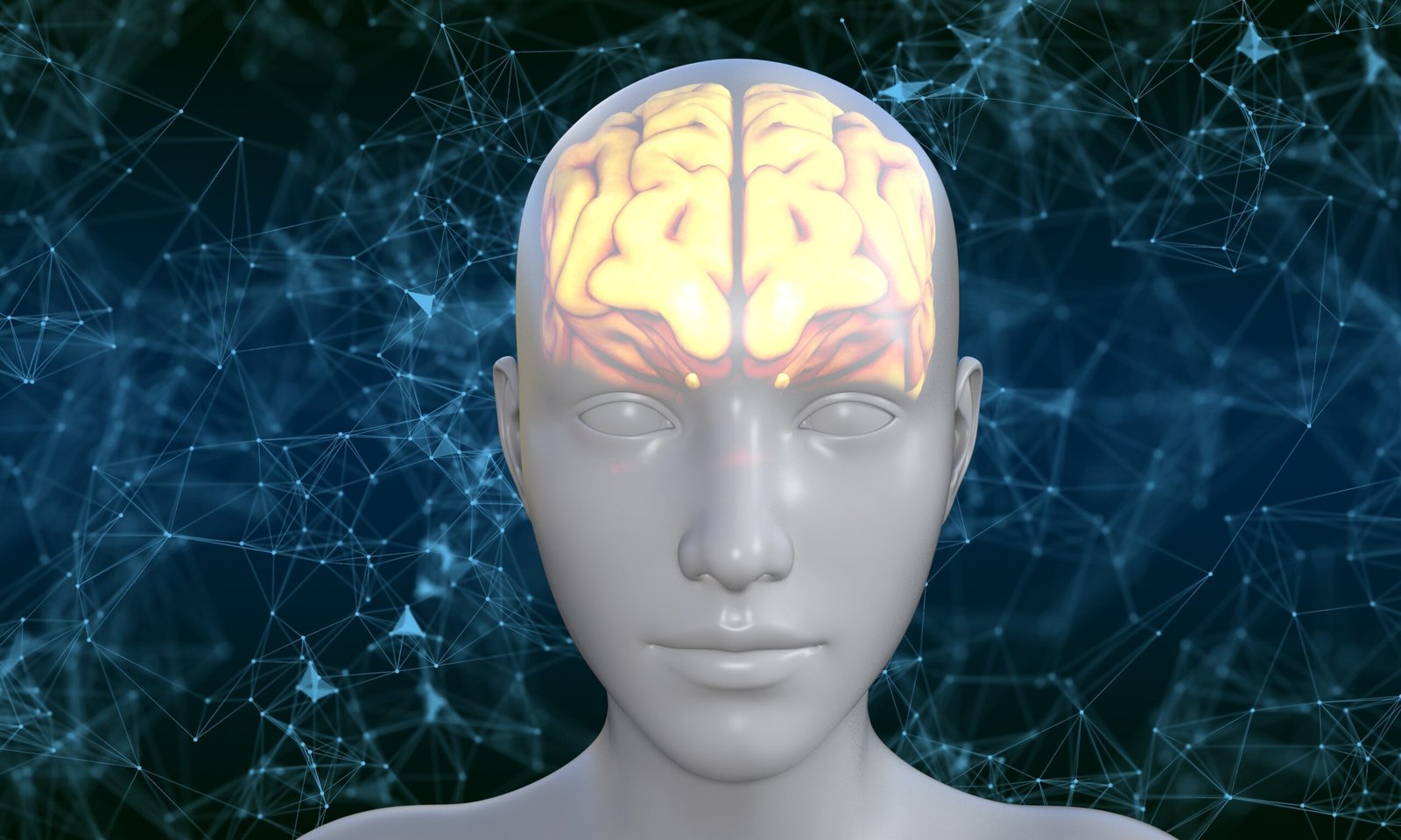

The human brain operates using extremely subtle electrical signals.
Every thought, memory, and heartbeat is coordinated by electrical signals measuring only 70 to 130 millivolts, which is less than one-tenth the voltage of a standard AA battery. Each neural spike consumes between 0.3 and 100 picojoules of energy, where a picojoule equals one trillionth of a joule.
This remarkable efficiency contributes to the brain’s status as the most powerful computing system known, capable of processing extraordinary complexity with minimal energy consumption.
For the first time, engineers have developed artificial neurons that precisely match biological neurons in signal amplitude, energy consumption, timing, and frequency response.
This research, published in Nature Communications by a team from the University of Massachusetts Amherst and MIT, represents a significant advancement in neuromorphic electronics, a field focused on developing computers that emulate brain function.
Why “Close Enough” Wasn’t Good Enough
The disparity between artificial and biological neurons has long posed a significant challenge, one that researchers have often underestimated.
Previous artificial neurons operated at voltages of at least 0.5 volts. Biological neurons fire at 70–130 millivolts. That’s roughly a four- to sevenfold mismatch in signal strength alone. In engineering terms, that gap means artificial neurons can’t be seamlessly connected to real biological tissue without translation layers, amplifiers, and complex circuitry in between.
This mismatch can be likened to a conversation in which one participant speaks at a normal volume while the other uses a megaphone; communication is possible but inefficient and prone to information loss. Achieving integrated bioelectronics requires that both artificial and biological components operate at compatible signal levels.
Beyond voltage, energy was another mismatch. Standard artificial neurons consume far more energy than biological neurons, which matters enormously for implantable devices that need to operate safely inside or near living tissue without generating heat or triggering chemical reactions.
The University of Massachusetts team, led by Jun Yao, aimed to address both challenges simultaneously, thereby enabling broader advancements in the field.
The Secret Ingredient: Protein Nanowires From a Microbe
The key to this breakthrough is a material that sounds more like science fiction. The breakthrough was enabled by protein nanowires derived from the microbe Geobacter sulfurreducens, a material whose properties were previously considered more characteristic of science fiction than engineering. al wires just 2 to 3 nanometers in diameter that it uses to conduct electricity during its natural metabolic processes. These nanowires are extraordinarily stable, engineered by evolution to function reliably in harsh outdoor environments.
When the researchers incorporated these protein nanowires into a specially designed memristor, a component whose resistance varies with its usage history, analogous to synaptic plasticity in the brain, they achieved a significant advancement. Both values sit squarely within biological ranges. The off-state resistance (~200 megaohms) closely matched typical cell membrane resistance. And across 1,000 consecutive test cycles, the memristor performed with exceptional consistency and stability, exceeding the performance of previous bio-amplitude memristors.
This reliable, ultra-low-voltage memristor served as the foundation for the artificial neuron. This stable, ultra-low-voltage memristor served as the foundational component for the artificial neuron. They built a complete artificial neuron by pairing it with simple circuit components: a capacitor and a resistor.
This configuration produced a device capable of replicating the full cycle of biological neuronal firing with high fidelity:
Integration: When small electrical pulses arrive (mimicking incoming signals from other neurons), the memristor’s internal structure slowly builds up just like charge accumulates in a real neuron before it fires.
Depolarization: Once enough charge accumulates and the threshold is crossed, the memristor rapidly switches on, causing the capacitor to charge quickly, mimicking the sudden rush of sodium ions into a biological neuron during an action potential.
Repolarization: The charged capacitor immediately pushes back against the memristor, switching it off. Then the capacitor slowly discharges through the resistor, mimicking the flow of potassium ions out of a cell to reset it. A refractory period, the brief window when a neuron can’t fire again, emerges naturally from this process. The output spikes achieved amplitudes up to 120 millivolts, durations of 1 to 5 milliseconds, and energy consumption as low as 0.2 picojoules per spike, all within biological ranges. This represents the first artificial neuron in which both voltage and energy are simultaneously confined to biological parameters.
The device also exhibits distinct responses to various input patterns, such as tonic firing, burst firing, and adapting signals, mirroring the behavior of biological neurons and indicating potential for advanced computing networks.
Teaching the Neuron to Respond to Chemistry
Real neurons don’t just process electrical signals. They’re also tuned and modulated by chemical-biological neurons; not only electrical signals, but also chemicals such as neurotransmitters (e.g., dopamine) and ions (e.g., sodium) that influence neuronal excitability. This process, known as neuromodulation, is fundamental to the brain’s capacity for adaptation, learning, and responses to the environment. By connecting a resistor to a chemical sensor, they created a neuron whose firing rate responds to the surrounding chemical environment. Two demonstrations stand out:
Sodium sensing: As the concentration of sodium ions increased within the physiological range found in the human body, the artificial neuron’s firing rate increased correspondingly, exactly as biological neurons do when extracellular sodium rises.
Dopamine sensing: Dopamine has a complex, dual role in the brain. At low concentrations, it excites neurons; at high concentrations, it can suppress them. The team built a graphene-based sensor that produces the same ambipolar (both excitatory and inhibitory) response, and the artificial neuron’s firing rate followed the same up-then-down pattern seen in biology. Importantly, the chemically sensitive system operates at the same ultralow energy levels as the original artificial neuron. Its overall energy consumption is at least 100 times lower than that of previously reported chemical artificial neurons. The artificial neurons.
Connecting to a Living Heart Cell
The most compelling demonstration presented in the study involves directly connecting the artificial neuron to a living biological cell, highlighting its potential for future medical applications. A biological cell.
The team used human embryonic stem cell-derived cardiomyocytes, heart muscle cells that fire electrical action potentials and contract rhythmically, much like neurons. They embedded an ultra-flexible graphene-mesh sensor into a 3D cardiac microtissue, enabling them to record the electrical activity of individual hearts. These electrical signals were subsequently transmitted to the artificial neuron. The artificial neuron.
When the heart cells were beating at their normal resting rate (~0.4 Hz), the artificial neuron remained silent; the input signals were too slow and spaced too far apart to cross the memristor’s integration threshold.
When the tissue was treated with norepinephrine, a drug used to treat heart failure that increases heart rate, the beating rate increased to ~0.6 Hz. That small change in frequency was enough to shift the memristor’s dynamics from leaking to accumulating, causing the artificial neuron to fire.
The artificial neuron detected the drug-induced change in the biological cell’s state in real time, without the need for intermediary computational processing.
The researchers further demonstrated that, when provided with emulated inputs from clinical recordings of arrhythmia (a dangerous irregular heartbeat), the artificial neuron accurately identified the abnormal pattern. This finding suggests that artificial neurons could function as real-time interpreters of biological cell states, representing a novel class of biosensors.
What This Actually Means
This research is situated at the intersection of neuromorphic computing, bioelectronic medicine, and materials science, advancing all three fields concurrently.
In the near term, the most immediate applications are anticipated in bioelectronic interfaces, which connect electronic devices to the nervous system or other electroactive tissues for purposes such as monitoring, stimulation, or therapy. Current devices, including cochlear implants, deep brain stimulators, and cardiac pacemakers, are limited by signal mismatch issues that this research addresses. Devices capable of operating at biological signal levels could be smaller, safer, more energy-efficient, and more precisely targeted.
In the longer term, the vision encompasses neuromorphic chips that process information with the energy efficiency and adaptability characteristic of biological computation. This includes integrating chemical sensing into neuromorphic systems and developing interfaces that enable direct, cellular-level communication between artificial and biological neurons. The protein nanowires central to this device are notable as a sustainably produced biological material that outperforms engineered alternatives in advanced electronics applications. This work is still preliminary. Scaling these individual demonstrations into practical systems will require significant further engineering. But the proof of concept is now established: artificial neurons can be built that are, in the ways that matter most, genuinely equivalent to the real thing.