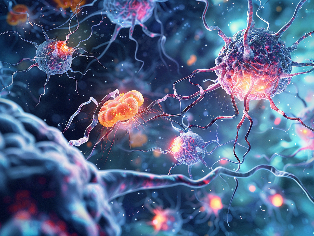

Millions of people across the world are afflicted with Parkinson diseases (PD) and the resultant aspect includes tremors, stiffness and progressive motor and cognitive impairments. The central component of this neurodegenerative disease is a protein known as a-synuclein (a-Syn). Although a-Syn is supposed to aid in the process of the communication between nerve cells, its mis folded version can form toxic oligomers (aSOs). Such minute and unstable proteinaceous aggregates are much more dangerous than the larger fibrils that have been identified as a hallmark of PD long before them.
The exact mechanism of action of ASOs damaging neurons by interfering with the finely tuned membranes that safeguard the brain cells and arrange the brain cells had remained a mystery although researchers had a hunch over the years that this is how the ASOs cause brain damage. However, there has been a breakthrough study by Brochner et al. (2025) that has revealed a more intricate image of the manner in which aSOs engage with membranes to create small holes that discharge molecules and destabilize cells.
What are a-Synuclein Oligomers?
The natural shapes of proteins are accurately folded 3D, whereas a-synuclein is an intrinsically disordered protein, i.e. it folds into various shapes. It may misfold and oligomerize under stress. Off-pathway oligomers are trapped in a toxic state unlike the on-pathway oligomers which develop into fibrils.
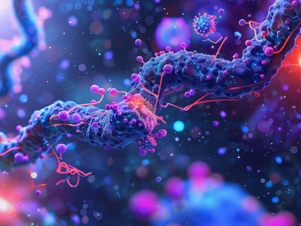
These aSOs have now been regarded as important contributors to PD toxicity since they directly interfere with cell membranes undermining their integrity and predisposing them to neuronal dysfunction.
The Three Stage Model of Membrane Damage.
The new study incorporated the powerful single-vesicle analysis workstation which enables the scientists to observe the interaction of single aSOs on artificial membranes in real time. Through the observation of over half a million interactions between vesicles and oligomers, the team found a three-step model of membrane perturbation.
Membrane Recruitment
Initially, aSOs bind to the exterior surface of cell membranes and particularly those that are negatively charged with the more abundant phosphatidylglycerol (PG) or phosphatidylserine (PS). Curved membranes (through synaptic vesicles) favor this first step.
Partial Pore Insertion
In the binding process, aSOs penetrate the lipid bi-layer partially. This step destabilizes the membrane but does not form a complete pore yet.
Full Pore Formation
Lastly, aSOs are completely embedded within the membrane creating dynamic pores, which allow small molecules to enter. Larger sized molecules such as dextran cannot cross- this shows that they are selective nanoscale channels and not random ruptures.
Notably, the formation of pore and recruitment occurs independently. Although binding is most successful on curved membranes, full pore formation is easier on flatter membranes as in the case of mitochondria.
Why is This Important to Parkinson?
Cell membranes do not simply form barriers. They control communication, metabolism and survival. In cases where pores develop out of control, ions and small molecules will leak out causing energy failure, oxidative stress, and eventual death of neurons.
The paper has also pointed out that:
The charge of the membrane is important: Negatively charged lipids increase the chance to form the pore significantly.
Curvature has a role: Recruitment is simpler on curved membranes, whereas stability of pores is stronger on flatter ones.
Dynamic cycling is observed: aSOs will be able to repeatedly alternate the partial and full pore state, which will subject cells to continuous stress.
Combined, these findings can bring a better understanding of how aSOs cause havoc within the brain and why they can serve as potential therapeutic targets.
Nanobodies: Little Wonders in the War on Toxic Oligomers
Another interesting thing about the research is the application of nanobodies (NBs)- small fragments of the antibodies that are camelid in nature. Two nanobodies were tested and had dissimilar influences on aSO pore formation:
NB1 surprisingly enhanced the activity of the pores, oligomers became more active.
NB2 decreased pore production, and it has potential as an agent of protection.
This is an indicator that that in the future, carefully engineered nanobodies or other such ligands may be used to block aSO pores to slow or prevent neuronal injury in PD.
Future Perspectives
The single vesicle assay that was designed in this research is not merely one of the tools of the research but a possible drug discovery platform. Using the screening of effects of various lipids, ligands or small molecules on aSO behavior scientists can quickly find candidates that stabilize membranes or prevent pore formation.
In addition, the results can be generalized to other disorders other than Parkinson. Toxic oligomers (e.g. tau or amyloid-b) are also involved in other neurodegenerative diseases which might interplay with membranes in the same manner.
Conclusion
This groundbreaking study illuminates the molecular mechanism of attack of a-synuclein oligomers: recruit, insert, and punch holes in neuronal membranes. Discovery of this three-step model has given scientists a guide on how to come up with treatments that stop or inhibit the creation of the pore.
In the case of patients of the condition known as Parkinson, these findings offer some hope that the next generations of treatment will be able to save the brain cells, as the processes of membrane disruption are going to be addressed at the earliest stage possible.
Reference
Brochner, B. V., Zhang, X., Nielsen, J., Kjems, J., Otzen, D. E., & Malle, M. G. (2025). Single vesicle Tracking of α-Synuclein Oligomers Reveals Pore Formation by a Three-Stage Model. ACS Nano. https://doi.org/10.1021/acsnano.5c04005