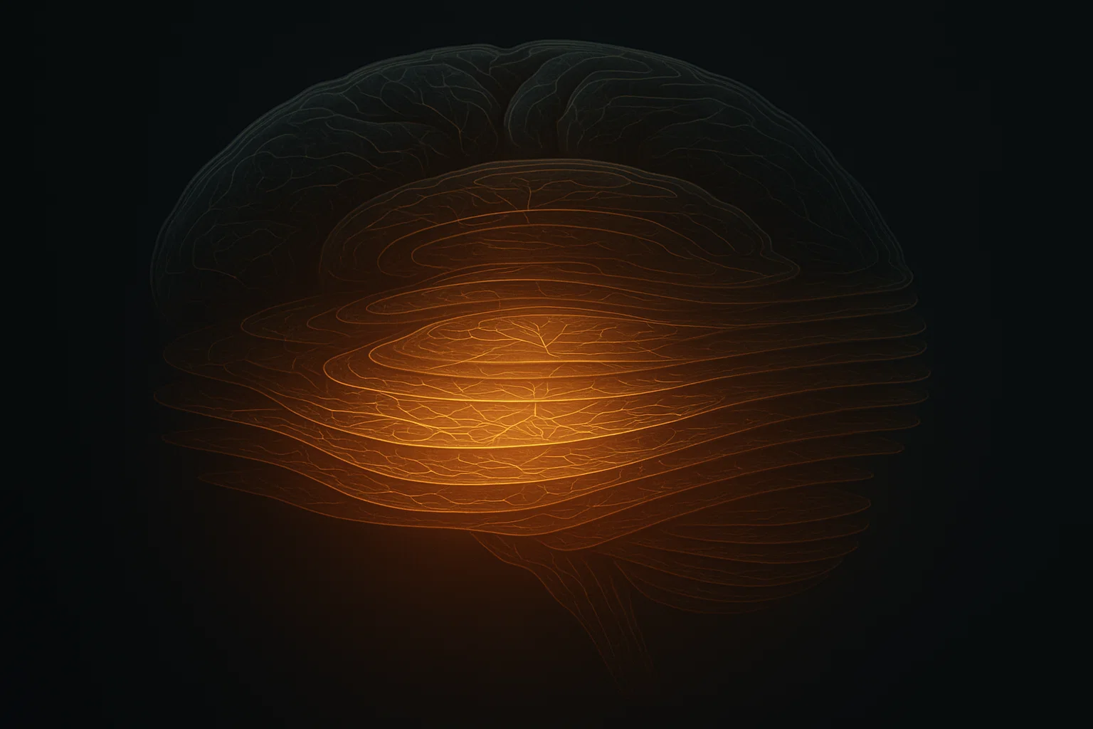

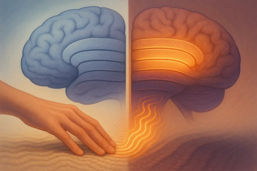
Our brains are similar to complex maps, where in several layers are dedicated to various duties receiving, sending, and so on. This is a multi layered arrangement, found in animals very ancient. But how does aging manipulate these layers, particularly those of the senses? A team in that area breaks new ground by merging sophisticated imaging in humans and mice in a 2025 paper in Nature Neuroscience to inspect the primary somatosensory cortex the touch processing center. The results indicate particular layer changes that led toward explanations of age specific sensory hiccups, such as diminished touch, or reduced responsiveness.
Led by Esther Kuehn of Otto von Guericke University Magdeburg, the researchers scanned two groups of healthy adults younger (20-40) and older (60+). They applied ultra high field 7T MRI to look at layer parts exactly, as well as functional scanning when the finger is stimulated. In mice, the calcium imaging and histology monitored neuronal activity and structure throughout ages.
Expanded Input Layers: A Mark of Aging Adaptation?
Outstanding finding: The key input station, Layer IV, increases and becomes myelinated (lightning fast through fatty covering) in aging adults. This correlates with longer sensory signals the elderly have longer and broader brain response to touch. What happens is that brain is like the receiving dock in the brain, which can increase its size in a bid to be able to receive signals better, perhaps offsetting weaker ones elsewhere.
In humans, age related cortical thinning affects the deep layers (V-VI) most severely, in which the outputs are generated. However, there is increased overall myelination indicating more wiring. No compelling evidence of slowed inhibition (braking overexcitation) was found, which confunders certain theories of aging.
Human and The Mouse Models Mirror The Changes
The pattern shown by mice proved cross species. Older mice (18-24 months) exhibited increased afferent driven neuron activity in whisker cortex, which was compensated by an increase in parvalbumin a measure of inhibitory cells. Myelination varied dynamically: reaching the maximum at the middle age term, then declining in the deep layers during old age.
Amplified activity traces in aged mice were observed in the calcium imaging of live neuron conversation during flicks of whiskers. Histologically parvalbumin levels showed elevations, suggesting build up of inhibition to overcome excitation.
The most vulnerable were middle (II-III) and deep layers, which accorded with human data. This is indicative of evolutionary preservation: output/modulatory layers get hit by aging first.
Touch Point to Wider Brain Health
The altered sensory cortex may have a spill over effect on cognition hence, inadequate touch feedback may slow learning or movement. The enlargement of Layer IV may prolong the signals, helping adaptation, yet causing overload.
There is no evidence to reverse the inhibition hypotheses; rather, the balance of excitation/inhibition remains, but myelin adjustments.
Advantages of study design: Multimodal (structural/functional MRI, histology), cross specimen. Limits: Greatly limited samples (30-40 human subjects per group), emphasis on somatosensory global next?
Healthy Aging Key Takeaways
Not much can be done to stop the march of time under our skin, but physical activity promotes myelination; cognitive activity prevents layer loss. These perceptual sharpenings may occur through sensory training (e.g. textured puzzles).
Researchers: Deep layers are the target of such interventions as myelin improving drugs.
This paper highlights stratum-wise perceptions to the post comprehension of dysfunction in aging to degenerative diseases such as Alzheimer where sensory problems begin early.
As we get older we restructure our brains in a very smart way. Changes are embraced through healthy habits to maintain the senses.
References & Resources
Liu, P., Doehler, J., Henschke, J.U., et al. (2025). Layer-specific changes in sensory cortex across the lifespan in mice and humans. Nature Neuroscience. https://doi.org/10.1038/s41593-025-02013-1
Salat, D.H., et al. (2004). Thinning of the cerebral cortex in aging. Cerebral Cortex.