
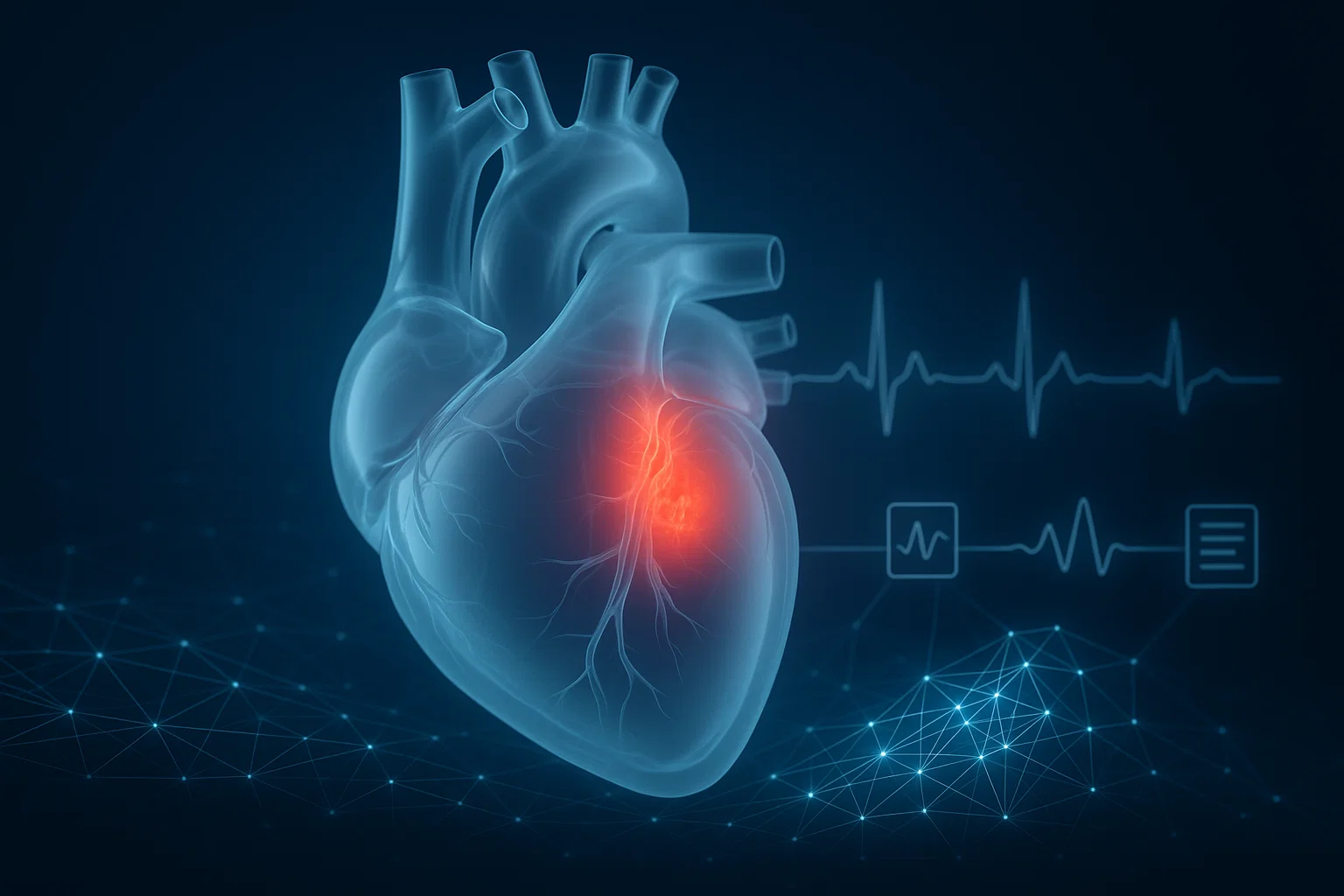

Artificial intelligence is helping scientists examine the heart in ways conventional medical assessments cannot. A system called MAARS combines cardiac MRI images, medical records, and imaging reports to estimate the risk of life-threatening ventricular arrhythmias in people with hypertrophic cardiomyopathy. Its early results are promising, but can AI truly improve sudden cardiac death prevention?
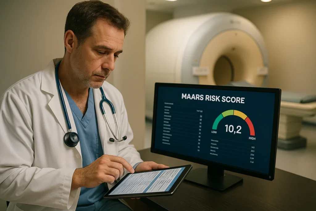
MAARS is an experimental artificial intelligence system designed to estimate the risk of dangerous ventricular arrhythmias in patients with hypertrophic cardiomyopathy (HCM).
Changxin Lai and colleagues published the original study in Nature Cardiovascular Research on July 2, 2025, with researchers associated with Johns Hopkins University.
Researchers evaluated 839 patients, including 553 in the model-development cohort and 286 in an independent external-validation cohort.
MAARS achieved an AUROC of 0.89 in internal testing and 0.81 in external validation, demonstrating stronger risk discrimination than the conventional clinical tools evaluated in the study.
The system combines cardiac MRI, medical records, and imaging reports, allowing it to detect complex patterns associated with arrhythmia risk.
As of October 2026, MAARS remains a research-stage approach. Further prospective testing is needed to determine whether using it to guide medical decisions improves patient outcomes.
The Challenge of Predicting Sudden Cardiac Death
Sudden cardiac death is among the most serious complications that can occur in people with certain heart conditions.
It happens when the heart unexpectedly stops functioning effectively, often because of a dangerous electrical disturbance.
One condition associated with an increased risk is hypertrophic cardiomyopathy, commonly abbreviated HCM.
HCM is characterised by abnormal thickening of the heart muscle that cannot be explained solely by conditions such as high blood pressure.
Many people with HCM live for decades without experiencing life-threatening complications. Others may develop heart failure symptoms, atrial fibrillation, or dangerous ventricular arrhythmias.
The challenge for cardiologists is determining which patients are at greatest risk.
This decision matters because an implantable cardioverter-defibrillator (ICD) can identify and terminate certain life-threatening heart rhythms.
Yet ICD implantation is an invasive procedure with possible complications.
Doctors must therefore balance the benefits of preventing sudden cardiac death against the risks of implanting a device unnecessarily.
In July 2025, researchers introduced a new AI model called MAARS that showed promising results in distinguishing higher-risk and lower-risk patients.
The findings suggested that combining different types of medical information could improve risk assessment beyond conventional approaches.
What Is Hypertrophic Cardiomyopathy?
Hypertrophic cardiomyopathy is a disease in which the heart’s muscular walls become abnormally thick.
In many patients, the thickening prominently affects the interventricular septum, which separates the heart’s two main pumping chambers.
However, HCM can involve other regions of the heart muscle.
It is often inherited and can involve disease-causing changes in genes that encode proteins responsible for heart muscle contraction.
The condition has historically been estimated to affect about 1 in 500 people, although prevalence estimates vary by population and diagnostic criteria.
Common Symptoms
Some people with HCM experience:
- Shortness of breath, especially during physical activity.
- Chest discomfort or pain.
- Palpitations or a racing heartbeat.
- Dizziness or unexplained fainting.
- Reduced exercise tolerance.
Other individuals have few or no noticeable symptoms.
HCM is also associated with an increased risk of certain abnormal heart rhythms.
Importantly, an HCM diagnosis does not mean that sudden cardiac death is inevitable. Many patients have a favourable prognosis with appropriate evaluation and management.
Clinical reference: American Heart Association — 2024 Guideline for the Management of Hypertrophic Cardiomyopathy.
Why Can HCM Cause Dangerous Arrhythmias?
The heart depends on carefully coordinated electrical signals to maintain its pumping rhythm.
In HCM, several structural and cellular changes can increase vulnerability to abnormal electrical activity.
These include altered heart muscle organisation, abnormal electrical conduction, and areas of myocardial fibrosis.
The Role of Heart Muscle Scarring
Myocardial fibrosis refers to the accumulation of scar-like tissue within the heart muscle.
Fibrosis can disrupt normal electrical signal propagation.
Instead of travelling through the heart in an orderly manner, electrical impulses may encounter areas that slow or interrupt conduction.
Under certain circumstances, this can help create the conditions for dangerous ventricular arrhythmias.
Two important examples are:
Ventricular tachycardia: An abnormally fast heart rhythm originating in the ventricles.
Ventricular fibrillation: Chaotic electrical activity that prevents the ventricles from pumping blood effectively.
These arrhythmias can lead to sudden cardiac arrest if they are not terminated promptly.
The distribution and extent of fibrosis are therefore important considerations when evaluating HCM-related arrhythmic risk.
How Doctors Currently Estimate Sudden Cardiac Death Risk
Current clinical evaluation considers multiple factors rather than relying on a single test.
Depending on the patient, cardiologists may assess:
- Previous cardiac arrest or sustained ventricular arrhythmias.
- A family history of premature HCM-related sudden death.
- Unexplained fainting that may indicate an arrhythmia.
- Marked thickening of the heart muscle.
- Episodes of nonsustained ventricular tachycardia.
- Reduced left ventricular ejection fraction.
- A left ventricular apical aneurysm.
- The extent of myocardial fibrosis seen on cardiac MRI.
Clinical guidelines and risk calculators help clinicians estimate the likelihood of serious complications.
The 2024 American Heart Association and American College of Cardiology guideline recommends systematic sudden cardiac death risk assessment at initial evaluation and every one to two years thereafter in adults with HCM.
However, risk estimation is imperfect.
Some people who appear to be at relatively low risk may experience serious events.
Others classified as higher risk may never develop a life-threatening arrhythmia.
The goal of improved prediction is not to replace clinical judgment, but to provide more reliable information for decisions about monitoring and preventive treatment.
What Is MAARS?
MAARS stands for Multimodal Artificial Intelligence for Ventricular Arrhythmia Risk Stratification.
It is a deep-learning system designed to analyse several types of clinical information simultaneously.
Changxin Lai and colleagues conducted the research, including researchers working with Natalia A. Trayanova at Johns Hopkins University.
Their study was published on July 2, 2025, in Nature Cardiovascular Research.
Original research: Lai et al. — Multimodal AI to forecast arrhythmic death in hypertrophic cardiomyopathy.
Free full-text study: PubMed Central — PMC12259465.
Unlike conventional models that use a limited set of predefined clinical measurements, MAARS analyses detailed patterns across multiple data sources.
These include electronic health records, cardiac imaging reports, and contrast-enhanced cardiac magnetic resonance images.
The model uses transformer-based neural networks, a type of AI architecture capable of learning complex relationships within data.
Its central innovation is integrating medical information that might otherwise be examined separately.
How Does MAARS Work?
MAARS combines three major information sources.
1. Electronic Health Records
The model analyses clinical variables extracted from patient records.
These may include demographic characteristics, clinical history, and other established risk-related measurements.
Such information provides context about the patient and their cardiovascular condition.
However, medical records alone may not capture the heart’s full structural complexity.
2. Echocardiogram and Cardiac MRI Reports
Echocardiography uses ultrasound to evaluate the heart’s structure and function.
Cardiac MRI provides additional information about heart muscle thickness, ventricular function, and tissue characteristics.
Reports from these examinations contain measurements that clinicians already use in HCM assessment.
MAARS processes relevant numerical information from these imaging reports as additional evidence.
3. Contrast-Enhanced Cardiac MRI Images
One of MAARS’s most distinctive features is its direct analysis of cardiac MRI images.
Specifically, the researchers used a technique called late gadolinium enhancement, or LGE.
This technique can reveal patterns associated with myocardial fibrosis.
Traditional clinical assessment may consider the amount of visible fibrosis.
MAARS can also learn from spatial patterns within the MRI images.
The model may therefore identify scar-distribution relationships that are difficult to capture with a small number of conventional measurements.
Combining the Information
MAARS processes these data sources through specialised neural-network branches.
A fusion component then combines the information to produce a risk estimate.
The model also incorporates methods designed to help researchers understand which features contribute to its predictions.
This is sometimes called explainable artificial intelligence.
Nevertheless, an explanation generated by a machine-learning model does not automatically establish that the identified features cause dangerous arrhythmias.
The 2025 MAARS Study: What Did Researchers Find?
The investigators evaluated MAARS using two independent groups of patients with HCM.
The first cohort came from Johns Hopkins Hospital in Maryland.
The second came from Sanger Heart & Vascular Institute in North Carolina.
Together, the two cohorts included 839 patients.
| Study group | Number of patients | Purpose |
|---|---|---|
| Johns Hopkins Hospital | 553 | Model development and internal cross-validation |
| Sanger Heart & Vascular Institute | 286 | Independent external validation |
| Total | 839 | Combined research population |
The study evaluated MAARS’s ability to distinguish patients who experienced serious ventricular arrhythmia events from those who did not.
The primary endpoint included sustained ventricular tachycardia or ventricular fibrillation associated with appropriate defibrillator treatment.
This distinction is important: the study did not simply measure whether the model could predict a documented death in every case.
MAARS Showed Stronger Risk Discrimination
The researchers assessed performance using the area under the receiver operating characteristic curve (AUROC).
An AUROC of 0.5 represents no better discrimination than chance, while a value of 1.0 represents perfect discrimination in the evaluated dataset.
MAARS achieved:
| Evaluation | MAARS AUROC |
|---|---|
| Internal cross-validation | 0.89 |
| External validation | 0.81 |
The model performed better than the conventional clinical risk-assessment approaches evaluated in the same study.
However, the decrease from 0.89 internally to 0.81 externally also shows why testing a model in different populations is essential.
An AUROC of 0.89 does not mean the system predicts an individual patient’s outcome with 89% certainty or prevents 89% of sudden deaths.
It describes how well the model distinguished patients with and without the evaluated events across the study population.
How Did MAARS Compare With Conventional Risk Assessment?
The researchers compared MAARS with established clinical approaches, including American and European guideline-based methods and the HCM Risk-SCD calculator.
The models were evaluated retrospectively using the study’s available clinical information.
The results were:
| Risk assessment approach | Internal AUROC | External AUROC |
|---|---|---|
| MAARS | 0.89 | 0.81 |
| Conventional approaches evaluated | Approximately 0.54–0.62 | Approximately 0.51–0.59 |
MAARS showed substantially better discrimination in the two research datasets.
These findings are encouraging, but the comparison needs context.
The study used retrospective data, and the guideline-based approaches were compared using the variables and definitions available to the investigators.
Clinical guidelines are broader decision frameworks, not exclusively prediction algorithms.
The study also compared MAARS with clinical tools available at the time of analysis, including earlier versions of certain guidelines.
Therefore, the results should not be interpreted to mean that existing guideline-based care is ineffective or should be abandoned.
Instead, MAARS demonstrates that combining detailed imaging with clinical data may improve how researchers estimate arrhythmic risk.
Why Cardiac MRI Scar Patterns Matter
Cardiac magnetic resonance imaging is particularly valuable for assessing the heart’s structure and tissue characteristics.
Late gadolinium enhancement can identify areas associated with replacement fibrosis.
In HCM, extensive fibrosis is linked to an increased risk of serious ventricular arrhythmias.
Clinicians already consider the extent of LGE when assessing risk.
However, cardiac scarring is not always distributed uniformly.
Some patients have localised regions of fibrosis, while others have more complex patterns involving different parts of the ventricular wall.
A machine-learning model may identify subtle relationships between these patterns and clinical outcomes.
This is one reason MAARS may offer more information than conventional approaches based only on broad clinical measurements.
The importance of combining imaging modalities was also emphasised in the 2025 European Association of Cardiovascular Imaging consensus on HCM.
Further reading: European Association of Cardiovascular Imaging consensus on multimodality imaging in HCM.
Could MAARS Help Doctors Make Better ICD Decisions?
An implantable cardioverter-defibrillator is a small electronic device that detects and treats dangerous heart rhythms.
For selected high-risk patients with HCM, an ICD can provide life-saving protection.
However, implantation involves a procedure and potential complications, including infection, inappropriate shocks, lead problems, and the need for future device replacement.
Accurate risk assessment is therefore essential.
MAARS could potentially help clinicians identify people whose risk may be underestimated by conventional methods.
It could also help provide additional information when the decision to implant an ICD is uncertain.
For example, a future clinically validated system might support discussions between patients and cardiologists by showing how imaging and clinical features contribute to an individualised risk estimate.
But the original MAARS study did not test whether using its predictions to guide ICD implantation improves survival.
It also did not show that MAARS-based decisions reduce unnecessary procedures.
The system’s potential to improve ICD selection remains a hypothesis requiring prospective clinical evaluation.
Can MAARS Explain Its Predictions?
One major concern about AI in healthcare is interpretability.
Complex neural networks may generate predictions without making their internal reasoning easy for clinicians to understand.
This is especially concerning when medical decisions carry serious consequences.
The MAARS researchers tried to address this problem using attention-based visualisation and feature-attribution methods.
These techniques can indicate which input features or image regions influence a prediction.
The researchers also used Shapley values to analyse the contributions of certain clinical variables.
This approach may help clinicians assess whether a model’s output aligns with known clinical information.
However, these interpretation techniques have limitations.
They describe features associated with a model’s prediction, rather than necessarily identifying the biological cause of an arrhythmia.
Before clinical adoption, researchers must determine whether the explanations are sufficiently reliable, stable, and understandable to support real medical decisions.
What Research After 2025 Adds to the Picture
The original MAARS study helped demonstrate how multiple data types could be combined for cardiac risk assessment.
Related research has continued to explore other ways AI might improve HCM evaluation.
September 2025: Detecting Heart Scarring From an ECG
A study published in Scientific Reports on September 30, 2025, investigated an explainable AI system called XplainScar.
Unlike MAARS, which combines cardiac MRI and multiple clinical data sources, XplainScar was developed to identify left ventricular scar-related information using standard 12-lead electrocardiograms.
The researchers used retrospective data from 748 patients with HCM across two institutions.
The findings suggested that ECG-based AI might eventually help identify patients who need further evaluation for myocardial fibrosis.
This approach is potentially important because ECG testing is generally less expensive and more widely accessible than cardiac MRI.
However, XplainScar does not replace MAARS and was designed for a different prediction task.
The study evaluated AI-based scar detection rather than establishing improved prevention of sudden cardiac death.
Original research: Explainable artificial intelligence identifies and localises left ventricular scar in hypertrophic cardiomyopathy using 12-lead electrocardiogram.
2026: Continued Interest in Multimodal Cardiac Imaging
Research and clinical reviews published in 2026 have continued emphasising the importance of combining echocardiography, cardiac MRI, and other diagnostic information in HCM.
A 2026 review, Multimodality Imaging in Hypertrophic Cardiomyopathy, discusses the value of cardiac imaging for diagnosis, assessment of myocardial fibrosis, risk stratification, and treatment planning.
This broader research supports the scientific rationale for multimodal approaches.
However, it should not be confused with an independent prospective validation of MAARS itself.
Further reading: Multimodality Imaging in Hypertrophic Cardiomyopathy (2026).
Has MAARS Been Adopted in Hospitals by October 2026?
The available published evidence supports MAARS as a promising research-stage prediction model.
The original investigators demonstrated performance through internal cross-validation and independent retrospective external validation.
However, those findings do not establish routine clinical deployment.
As of October 30, 2026, I could not verify completed prospective evidence demonstrating that MAARS-guided clinical decisions reduce sudden cardiac death, improve survival, or safely reduce ICD implantation.
A research model can perform well in a retrospective dataset but not produce the same results in everyday clinical practice.
Clinical implementation would require further testing to establish whether predictions remain reliable when patients are evaluated prospectively under real-world conditions.
Researchers would also need to determine how clinicians should respond to a high-risk or low-risk MAARS result.
Until such evidence becomes available, established clinical guidelines and specialist judgment remain central to HCM management.
What Are the Limitations of MAARS?
Several issues need to be addressed before MAARS could become a routine clinical decision-support system.
1. Retrospective Study Design
The original research relied on previously collected patient data.
Prospective studies are needed to assess whether MAARS performs reliably in patients evaluated in real time.
2. Relatively Small Numbers of Arrhythmic Events
Sudden cardiac death and sustained life-threatening ventricular arrhythmias are relatively uncommon compared with the total number of people living with HCM.
The Johns Hopkins cohort included 19 patients who experienced the study’s primary arrhythmic endpoint during follow-up.
Small event counts can increase uncertainty around predictive performance.
3. Limited Geographic Representation
The original research involved two U.S. medical centres.
Further evaluation in other healthcare systems and populations would be important before broad adoption.
4. Dependence on Advanced Imaging
MAARS uses contrast-enhanced cardiac MRI data.
These examinations may be expensive, require specialised equipment, and be less accessible in some regions.
Certain patients may also be unable to undergo particular MRI examinations or contrast-enhanced imaging.
5. Incomplete Time-to-Event Modelling
The original investigators acknowledged limitations involving detailed time-to-event information.
As a result, the study did not fully establish how risk predictions change over specific periods or how they should be translated into long-term treatment decisions.
6. Risk of False Positives and False Negatives
No prediction model is perfect.
A false-positive prediction could contribute to unnecessary anxiety or testing.
A false-negative prediction could create inappropriate reassurance.
These potential consequences must be considered before integrating AI predictions into clinical care.
7. Prediction Is Not Prevention
Even if a model identifies higher-risk patients more accurately, improved health outcomes depend on whether clinicians can use that information to choose effective interventions.
Prospective clinical studies must establish that this connection exists.
What Does MAARS Mean for Patients With HCM Today?
For patients currently living with HCM, MAARS represents a promising area of research rather than a replacement for established medical care.
People with HCM should continue to receive individualised follow-up from qualified cardiovascular professionals.
According to the 2024 AHA/ACC guideline, risk assessment may involve clinical history, electrocardiographic monitoring, echocardiography, and cardiac MRI when indicated.
For some patients, an ICD may be recommended or considered based on established risk markers.
The decision should involve careful discussion of potential benefits, complications, patient preferences, and the person’s overall clinical situation.
Patients should not change medication, stop follow-up appointments, or decline an ICD recommendation based on research reports about MAARS.
Anyone experiencing sudden collapse, severe chest pain, or other symptoms suggesting a cardiac emergency should seek immediate emergency medical assistance.
Conclusion: A Promising Advance in Precision Cardiology
The development of MAARS represents an important step in applying artificial intelligence to cardiovascular medicine.
By combining cardiac MRI images with electronic health records and diagnostic reports, researchers developed a model that can identify complex patterns associated with life-threatening ventricular arrhythmias in people with HCM.
The 2025 study demonstrated encouraging discrimination in both internal and independent external testing.
It also highlighted the potential value of analysing the spatial distribution of myocardial fibrosis, rather than relying exclusively on conventional clinical measurements.
Related AI research has continued to investigate how cardiac imaging and electrocardiographic signals might reveal additional information about heart disease.
However, the clinical implications must be interpreted carefully.
MAARS has demonstrated promising predictive performance, but it has not yet been proven to reduce sudden cardiac death or improve treatment decisions in routine clinical practice.
The next step is to determine whether this technology can safely and reliably help cardiologists make better decisions for individual patients.
If future prospective trials confirm clinical benefit, MAARS and similar systems could become valuable additions to precision cardiovascular care.
For now, its greatest contribution is demonstrating how artificial intelligence may help researchers understand cardiac risk more deeply.
References
1. Lai, C., Yin, M., Kholmovski, E. G., et al. (2025). Multimodal AI to forecast arrhythmic death in hypertrophic cardiomyopathy. Nature Cardiovascular Research, 4, 891–903.
https://doi.org/10.1038/s44161-025-00679-1
2. Lai et al. (2025). Free full-text version of the MAARS study. PubMed Central.
https://pmc.ncbi.nlm.nih.gov/articles/PMC12259465/
3. Ommen, S. R., et al. (2024). 2024 AHA/ACC/AMSSM/HRS/PACES/SCMR Guideline for the Management of Hypertrophic Cardiomyopathy. Circulation.
https://doi.org/10.1161/CIR.0000000000001250
4. American Heart Association (2024). Guideline for the Management of Hypertrophic Cardiomyopathy.
https://professional.heart.org/en/science-news/2024-guideline-for-the-management-of-hypertrophic-cardiomyopathy
5. Nezamabadi, K., et al. (2025). Explainable artificial intelligence identifies and localises left ventricular scar in hypertrophic cardiomyopathy using 12-lead electrocardiogram. Scientific Reports, 15, 33918.
https://doi.org/10.1038/s41598-025-09282-7
6. European Association of Cardiovascular Imaging (2025). Role of multi-modality cardiac imaging in the management of patients with hypertrophic cardiomyopathy: A clinical consensus statement. European Heart Journal – Cardiovascular Imaging.
https://doi.org/10.1093/ehjci/jeaf282
7. Multimodality Imaging in Hypertrophic Cardiomyopathy (2026). Review of diagnostic and prognostic cardiac imaging approaches.
https://pmc.ncbi.nlm.nih.gov/articles/PMC13284785/
8. Johns Hopkins University (2025). AI Predicts Patients Likely to Die of Sudden Cardiac Arrest. Research announcement describing the MAARS project.
https://heartadvance.jhu.edu/2025/07/02/ai-predicts-patients-likely-to-die-of-sudden-cardiac-arrest/